. 2014 Sep;155(9):1861-1870.
doi: 10.1016/j.pain.2014.06.017. Epub 2014 Jun 25.

Human sensory neurons: Membrane properties and sensitization by inflammatory mediators

Affiliations

Human sensory neurons: Membrane properties and sensitization by inflammatory mediators

Steve Davidson et al. Pain. 2014 Sep.

Abstract

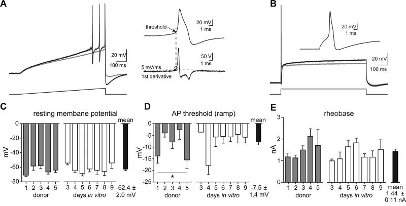
Biological differences in sensory processing between human and model organisms may present significant obstacles to translational approaches in treating chronic pain. To better understand the physiology of human sensory neurons, we performed whole-cell patch-clamp recordings from 141 human dorsal root ganglion (hDRG) neurons from 5 young adult donors without chronic pain. Nearly all small-diameter hDRG neurons (<50 μm) displayed an inflection on the descending slope of the action potential, a defining feature of rodent nociceptive neurons. A high proportion of hDRG neurons were responsive to the algogens allyl isothiocyanate (AITC) and ATP, as well as the pruritogens histamine and chloroquine. We show that a subset of hDRG neurons responded to the inflammatory compounds bradykinin and prostaglandin E2 with action potential discharge and show evidence of sensitization including lower rheobase. Compared to electrically evoked action potentials, chemically induced action potentials were triggered from less depolarized thresholds and showed distinct afterhyperpolarization kinetics. These data indicate that most small/medium hDRG neurons can be classified as nociceptors, that they respond directly to compounds that produce pain and itch, and that they can be activated and sensitized by inflammatory mediators. The use of hDRG neurons as preclinical vehicles for target validation is discussed.

Keywords: Bradykinin; Dorsal root ganglia; Human; Itch; Nociception; Pain; Sensitization.


Figures

Figure 1
Figure 1. Physical characteristics and capacitance of hDRG neurons
A) Phase-contrast images depicting dissociated hDRG neurons illustrating neurons that are suitable for patch-clamp recordings. Scale bar = 50 μm. B) Histogram summarizing the range of hDRG soma diameters from a subset of recorded neurons. Diameters were determined using a calibrated ocular eyepiece. C) Histogram of whole-cell capacitance from recorded hDRG neurons. D) Summary graph of the whole-cell capacitance from all donors and across time in vitro. Number of neurons indicated in parentheses: 3 DIV (6), 4 DIV (21), 5 DIV (16), 6 DIV (35), 7 DIV (21), 8 DIV (11), 9 DIV (19). n.s., not significantly different. E) Plot of whole-cell capacitance vs. soma diameter indicating a linear relationship between these two measurements. R2 = 0.336; Y=4.84x + 23.0 (p<0.0001). Dotted lines represent 95% confidence interval.
Figure 2
Figure 2. Excitability of hDRG neurons
A) Action potentials evoked by a 500 ms ramp current injection protocol. Gray trace depicts membrane voltage during a subthreshold step, with the subsequent sweep in the series of increasing current injections eliciting action potentials (black trace). Current steps were increased 50-100 pA per sweep. AP threshold was taken as the membrane voltage at the point in which the 1st derivative waveform of the first action potential crossed 5 volts/second (dashed line). B) Rheobase was established from an 800 ms step current protocol, increased in 50-100 pA increments. The gray trace illustrates a subthreshold depolarization, with the following sweep eliciting an action potential at the start. A majority of hDRG (70%) fired an action potential during the initial rising step of the current injection. C) Summary graph of the resting membrane potential across donors and time in culture, which was not significantly different. D) Summary of the action potential threshold across donors and time in culture, determined by ramp current injections shown in panel A. Action potential thresholds were variable by donor (*p<0.05, 1-way ANOVA) although no post-hoc comparisons between any groups were significant (Tukey's Multiple Comparison Test). Threshold did not differ by days in vitro. E) Graph of rheobase (step current thresholds to elicit an AP), which did not differ by donor or days in vitro.
Figure 3
Figure 3. Waveform and shoulder analysis of hDRG
A) Phase-plane plots of hDRG neurons showing examples from cells with a large shoulder (top) and small shoulder (bottom). Boxed regions indicate the onset and offset of the shoulder, depicted by the slowing rate of membrane voltage change. B) Histogram showing a distribution of the range of shoulder areas. Inset shows an example action potential waveform and the method used for calculating the shoulder area. Shoulder size was approximated using the formula ½(height*base). C) Scatter plot of the shoulder areas relative to cell size or current injection threshold. Shoulder areas did not correlate with either measure. Linear fits of the data are shown; R2=0.013, p=0.475 (size); R2=0.003, p=0.715 (rheobase).
Figure 4
Figure 4. Human DRG exhibit a wide range of afterhyperpolarization decay kinetics that correlate with action potential widths
A) Example traces of action potentials evoked by step current injections in three different human DRG neurons that exhibited markedly different action potential widths and afterhyperpolarization kinetics. B) Distribution of the mean weighted tau values from fitting the AP afterhyperpolarization from 86 hDRG neurons. Tau values were determined from either single or biexponential fits of the voltage decay. C, D) Scatter plots showing a strong linear correlation between afterhyperpolarization tau values and action potential widths, but not whole-cell capacitance measurements. Linear fits of the data are shown; R2 = 0.432, p<0.0001 (panel C); R2=0.002, p=0.6989 (panel D).
Figure 5
Figure 5. Human DRGs are activated by chemical algogens and pruritogens
A-D) Voltage traces depicting responses to AITC (30 μM), ATP (100 μM), histamine (100 μM), and chloroquine (100 μM). Bath application of compounds, indicated by colored rectangles, resulted in action potential discharge in a subset of cells. The number of responsive neurons is indicated in parentheses next to each chemical. The insets depict the AP waveforms of the first action potential in response to chemical application. Dashed lines represent −60 mV.
Figure 6
Figure 6. Human DRG excitability is modulated by the inflammatory compounds bradykinin and PGE2
A) Five minute bath application of bradykinin (100 nM) and B) prostaglandin E2 (1 μM) produced action potential discharge in a subset of hDRG neurons. These traces are in the continued presence of the indicated inflammatory mediator. C, D) Voltage traces of action potential firing during threshold step current injections before (black traces), and after exposure to either bradykinin (red) or PGE2 (blue). Neurons often fired multiple action potentials at rheobase following application of either compound. E) Summary graph of the injected current threshold to elicit action potentials in neurons before (naïve) and after bradykinin application. Nearly every cell exhibited a lower rheobase after exposure to bradykinin. Paired t-test, ***p<0.001. F) Graph of average membrane voltage during electrically-evoked action potentials (threshold of 5 mV/ms), before (naïve, dark gray), after bradykinin (red) exposure. Chemically evoked action potentials had much lower thresholds than those evoked electrically (light gray); ****p<0.0001. G) Table summarizing the responses to bradykinin (subset of 27 total neurons exposed). Step current-evoked action potentials were delayed (>10 ms) in 33% of hDRG neurons. These “delayed-type” neurons exhibited significantly greater probability of displaying chemically-evoked ongoing activity. 78% of these neurons fired action potentials, whereas only 33% of cells that fired early action potentials exhibited bradykinin-evoked discharge. Fisher's exact test, p<0.05. H) Distribution and average AHP tau values for neurons grouped based on their response to bradykinin. Of the 27 cells exposed to bradykinin, 14 experienced transient depolarization with action potential discharge, while the other 13 cells fired action potentials. **p<0.01.

Comment in

References

    1. Anand U, Facer P, Yiangou Y, Sinisi M, Fox M, McCarthy T, Bountra C, Korchev YE, Anand P. Angiotensin II type 2 receptor (AT2 R) localization and antagonist-mediated inhibition of capsaicin responses and neurite outgrowth in human and rat sensory neurons. European journal of pain. 2013;17(7):1012–1026. - PMC - PubMed
    1. Anand U, Otto WR, Casula MA, Day NC, Davis JB, Bountra C, Birch R, Anand P. The effect of neurotrophic factors on morphology, TRPV1 expression and capsaicin responses of cultured human DRG sensory neurons. Neuroscience letters. 2006;399(1-2):51–56. - PubMed
    1. Anand U, Otto WR, Sanchez-Herrera D, Facer P, Yiangou Y, Korchev Y, Birch R, Benham C, Bountra C, Chessell IP, Anand P. Cannabinoid receptor CB2 localisation and agonist-mediated inhibition of capsaicin responses in human sensory neurons. Pain. 2008;138(3):667–680. - PubMed
    1. Bandell M, Story GM, Hwang SW, Viswanath V, Eid SR, Petrus MJ, Earley TJ, Patapoutian A. Noxious cold ion channel TRPA1 is activated by pungent compounds and bradykinin. Neuron. 2004;41(6):849–857. - PubMed
    1. Basbaum AI, Bautista DM, Scherrer G, Julius D. Cellular and molecular mechanisms of pain. Cell. 2009;139(2):267–284. - PMC - PubMed

Publication types

MeSH terms