

The zebrafish as a tool to identify novel therapies for human cardiovascular disease
- PMID: 24973746
- PMCID: PMC4073266
- DOI: 10.1242/dmm.016170
The zebrafish as a tool to identify novel therapies for human cardiovascular disease
Abstract
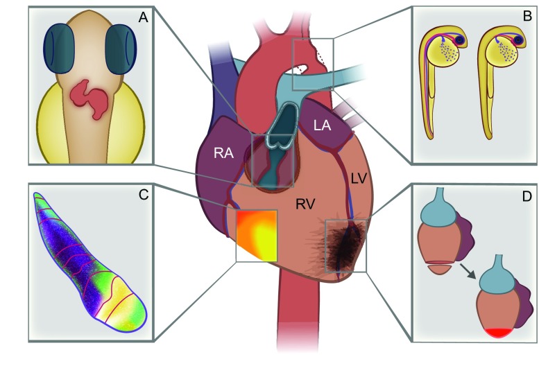
Over the past decade, the zebrafish has become an increasingly popular animal model for the study of human cardiovascular disease. Because zebrafish embryos are transparent and their genetic manipulation is straightforward, the zebrafish has been used to recapitulate a number of cardiovascular disease processes ranging from congenital heart defects to arrhythmia to cardiomyopathy. The use of fluorescent reporters has been essential to identify two discrete phases of cardiomyocyte differentiation necessary for normal cardiac development in the zebrafish. These phases are analogous to the differentiation of the two progenitor heart cell populations in mammals, termed the first and second heart fields. The small size of zebrafish embryos has enabled high-throughput chemical screening to identify small-molecule suppressors of fundamental pathways in vasculogenesis, such as the BMP axis, as well as of common vascular defects, such as aortic coarctation. The optical clarity of zebrafish has facilitated studies of valvulogenesis as well as detailed electrophysiological mapping to characterize the early cardiac conduction system. One unique aspect of zebrafish larvae is their ability to oxygenate through diffusion alone, permitting the study of mutations that cause severe cardiomyopathy phenotypes such as silent heart and pickwick(m171), which mimic titin mutations observed in human dilated cardiomyopathy. Above all, the regenerative capacity of zebrafish presents a particularly exciting opportunity to discover new therapies for cardiac injury, including scar formation following myocardial infarction. This Review will summarize the current state of the field and describe future directions to advance our understanding of human cardiovascular disease.
Keywords: Cardiovascular; Drug discovery; Zebrafish.
© 2014. Published by The Company of Biologists Ltd.
Figures
References
-
- Beis D., Bartman T., Jin S. W., Scott I. C., D’Amico L. A., Ober E. A., Verkade H., Frantsve J., Field H. A., Wehman A., et al. (2005). Genetic and cellular analyses of zebrafish atrioventricular cushion and valve development. Development 132, 4193–4204 - PubMed
-
- Benson D. W., Silberbach G. M., Kavanaugh-McHugh A., Cottrill C., Zhang Y., Riggs S., Smalls O., Johnson M. C., Watson M. S., Seidman J. G., et al. (1999). Mutations in the cardiac transcription factor NKX2.5 affect diverse cardiac developmental pathways. J. Clin. Invest. 104, 1567–1573 - PMC - PubMed
Publication types
MeSH terms
Grants and funding
LinkOut - more resources
Full Text Sources
Other Literature Sources
Miscellaneous
