Review
. 2014 Jul;7(7):785-97.
doi: 10.1242/dmm.015594.

Macrophage-pathogen interactions in infectious diseases: new therapeutic insights from the zebrafish host model

Affiliations
Review

Macrophage-pathogen interactions in infectious diseases: new therapeutic insights from the zebrafish host model

Vincenzo Torraca et al. Dis Model Mech. 2014 Jul.

Abstract

Studying macrophage biology in the context of a whole living organism provides unique possibilities to understand the contribution of this extremely dynamic cell subset in the reaction to infections, and has revealed the relevance of cellular and molecular processes that are fundamental to the cell-mediated innate immune response. In particular, various recently established zebrafish infectious disease models are contributing substantially to our understanding of the mechanisms by which different pathogens interact with macrophages and evade host innate immunity. Transgenic zebrafish lines with fluorescently labeled macrophages and other leukocyte populations enable non-invasive imaging at the optically transparent early life stages. Furthermore, there is a continuously expanding availability of vital reporters for subcellular compartments and for probing activation of immune defense mechanisms. These are powerful tools to visualize the activity of phagocytic cells in real time and shed light on the intriguing paradoxical roles of these cells in both limiting infection and supporting the dissemination of intracellular pathogens. This Review will discuss how several bacterial and fungal infection models in zebrafish embryos have led to new insights into the dynamic molecular and cellular mechanisms at play when pathogens encounter host macrophages. We also describe how these insights are inspiring novel therapeutic strategies for infectious disease treatment.

Keywords: Burkholderia; Candida; Host-directed therapy; Infectious disease; Innate immunity; Leukocyte biology; Mycobacterium; Salmonella; Shigella; Staphylococcus.


Figures

Fig. 1.
Fig. 1.
Evasion of macrophage defense mechanisms by intracellular pathogens. Upon phagocytosis (1), the pathogens generally reside within phagosomal compartments where a plethora of microbicidal components cooperate in a multidirectional assault to the microbes (2). By transferring virulence factors, often via secretion systems (injectosomes), some pathogens can avoid the classical maturation steps of these compartments, creating a favorable niche for their intracellular growth (3, 4, 5). Fusion of the phagosome with endosomes and/or lysosomes can be blocked (6) and fusion with Golgi- and reticulum-like vesicles can be promoted (3), resulting in the formation of specialized replicative vacuoles (4, 5), in some cases also directed to non-lytic expulsion (7). Several intracellular pathogens are able to escape directly into the cytosol (8). Here, septin cages (9), galectin decoration (10), ubiquitylation (11) and specific routes of antimicrobial autophagy (12) are activated to capture the escapers and redirect them to lytic compartments. Additionally, ubiquitylation of microbial proteins (11) labels these for proteasomal degradation (13). Several intracellular pathogens can efficiently counteract this second line of intracellular defense and replicate freely within the cytosol (14), frequently also manipulating the cell cytoskeleton (15) to sustain their extrusion and dissemination to other host cells (16). Intracellular infections have profound influences also on a wide spectrum of host functions. Cell signaling pathways can be manipulated to modulate the host inflammatory response (17) and control gene expression (18). Some pathogens are also known to induce epigenetic modification of their host cells, leading to reprogramming (19). Some virulence factors directly impact the homeostatic mechanisms by interfering with normal mitochondrial functionality (20), membrane polarity and communication with the extracellular milieu (21). The ultimate possibility for the host to eradicate the infection is to initiate cell (pyroptotic, apoptotic or necroptotic) suicide programs (20, 22, 23, 24). However, the death mechanisms can also be modulated by pathogens, which can benefit from them by the induction of host damage, pathogen dissemination and the initiation of new replicative cycles.
Fig. 2.
Fig. 2.
In vivo imaging of macrophage responses to infection. (A) A 3-dpf Tg(mpeg1:Gal4-VP16/UAS-E1b:Kaede) zebrafish embryo showing the distribution pattern of macrophages (green). Random patrolling of macrophages is shown in supplementary material Movie 1. (B) Phagocytosis of M. marinum (Mm; green) injected into the subcutaneous area overlying a somite in a 2-dpf Tg(mpeg1:mCherry-F) embryo. The arrow points at a macrophage (red) in the process of phagocytosis between 47 and 60 minutes post-infection. The images are particulars and stills from supplementary material Movie 2 (10 to 60 minutes post-infection). (C) Macrophage-mediated dissemination of M. marinum infection. The white track represents the path of an infected macrophage migrating away from the infection focus. The images are stills and particulars from supplementary material Movie 3, which was taken from the same embryo as in B at a more advanced stage of infection (~8 to ~10 hours post-infection). (D) Partial acidification of phagocytosed M. marinum. Bacteria double-labeled with constitutive mCrimson and pH-sensitive green pHrodo are contained within subcellular compartments of macrophages, which are intensely labeled by the membrane-bound mCherry of the Tg(mpeg1:mCherry-F) line. White arrows point at bacteria in acidified compartments, where the pHrodo dye is activated. Yellow arrows point at bacteria in non-acidified compartments. Note that most of the intracellular mycobacteria are not acidified, consistent with the ability of this pathogen to counteract phagosome maturation. Macrophages were imaged in the yolk sac circulation valley 5 hours after injection of bacteria into the caudal vein at 2 dpf. Images in A–C were acquired with the Zeiss Observer 6.5.32 laser-scanning confocal, with 10× (A) or 20× (B,C) objectives. Images in D were acquired with Leica TCS SPE confocal with a 20× objective. Figures and movies were processed with ImageJ. The zebrafish transgenic lines Tg(mpeg1:Gal4-VP16/UAS-E1b:Kaede) and Tg(mpeg1:mCherry-F) were previously described in other reports (Ellett et al., 2011; Bernut et al., 2014). Scale bars: (A) 200 μm; (B,C) 25 μm; (D) 10 μm.
Fig. 3.
Fig. 3.
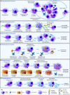
Models of intracellular infections in zebrafish. Schematic comparison of the infection phenotypes caused by different pathogens following intravenous injection in zebrafish embryos. (A) M. marinum can replicate within phagosomes and also escape into the cytosol. Eventually the host macrophages succumb to the infection, and the pathogen spreads to new macrophages that have been recruited through bacterial virulence mechanisms. This leads to the formation of granulomatous lesions. Occasionally, infected macrophages can egress from the primary granuloma and seed secondary granulomas. (B) S. typhimurium avoids the phagosomal defenses by inducing the formation of non-lytic compartments [Salmonella-containing vacuoles (SCVs)]. Upon replication, the pathogen induces pyroptotic cell death. Extracellular Salmonella continues to replicate. Damage- and pathogen-associated signals contribute to uncontrolled inflammation (‘cytokine storm’), which is rapidly fatal for the host. (C) B. cenocepacia can also replicate within intracellular compartments. Additionally, it can be non-lytically expelled from the host macrophages. Within the extracellular environment, the pathogen stimulates leukocyte aggregation and continues replication. The resulting uncontrolled bacteremia is the major cause of the fatal complications. (D) S. aureus is largely resistant to intracellular killing when phagocytosed by macrophages and leads to their necrotic death. By contrast, when phagocytosed by neutrophils, most of the pathogen can be efficiently neutralized. However, resistant clones occasionally emerge and expand within these cells. This ‘intraphagocyte niche’ is the reason of the monoclonality of focal staphylococcal abscesses. (E) S. flexneri can invade non-immune cells. Within these cells the pathogen escapes immediately into the cytosol and gains actin-based motility, by which it disseminates from cell to cell. Within macrophages, the pathogen can also escape from the phagosome, but here a more efficient cytosolic control partially combats the invader, delaying (although not avoiding) macrophage cell death. Neutrophils represent efficient scavengers for extracellular Shigella released from dying epithelial cells and macrophages but the infection is still rapidly lethal. (F) Phagocytosis of C. albicans by macrophages leads to a standoff phase, where the host does not degrade the pathogen, but its virulence is contained as it remains locked into a yeast form. The fungus can still slowly replicate and eventually is released. Extracellularly, the yeast can germinate and the resulting fast-replicating hyphae will invade the whole organism, leading to systemic infection.

Similar articles

Cited by

References

    1. Abdallah A. M., Gey van Pittius N. C., Champion P. A., Cox J., Luirink J., Vandenbroucke-Grauls C. M., Appelmelk B. J., Bitter W. (2007). Type VII secretion – mycobacteria show the way. Nat. Rev. Microbiol. 5, 883–891 - PubMed
    1. Adams K. N., Takaki K., Connolly L. E., Wiedenhoft H., Winglee K., Humbert O., Edelstein P. H., Cosma C. L., Ramakrishnan L. (2011). Drug tolerance in replicating mycobacteria mediated by a macrophage-induced efflux mechanism. Cell 145, 39–53 - PMC - PubMed
    1. Adams K. N., Szumowski J. D., Ramakrishnan L. (2014). Verapamil, and its metabolite norverapamil, inhibit macrophage-induced, bacterial efflux pump-mediated tolerance to multiple anti-tubercular drugs. J. Infect. Dis. [Epub ahead of print] 10.1093/infdis/jiu095 - DOI - PMC - PubMed
    1. Agnoli K., Schwager S., Uehlinger S., Vergunst A., Viteri D. F., Nguyen D. T., Sokol P. A., Carlier A., Eberl L. (2012). Exposing the third chromosome of Burkholderia cepacia complex strains as a virulence plasmid. Mol. Microbiol. 83, 362–378 - PubMed
    1. Agnoli K., Frauenknecht C., Freitag R., Schwager S., Jenul C., Vergunst A., Carlier A., Eberl L. (2014). The third replicon of members of the Burkholderia cepacia Complex, plasmid pC3, plays a role in stress tolerance. Appl. Environ. Microbiol. 80, 1340–1348 - PMC - PubMed

Publication types

MeSH terms