. 2014 Aug 5;393(1-2):129-42.
doi: 10.1016/j.mce.2014.06.010. Epub 2014 Jun 26.

Palmitoleic acid prevents palmitic acid-induced macrophage activation and consequent p38 MAPK-mediated skeletal muscle insulin resistance

Affiliations

Palmitoleic acid prevents palmitic acid-induced macrophage activation and consequent p38 MAPK-mediated skeletal muscle insulin resistance

Nicola A Talbot et al. Mol Cell Endocrinol. .

Abstract

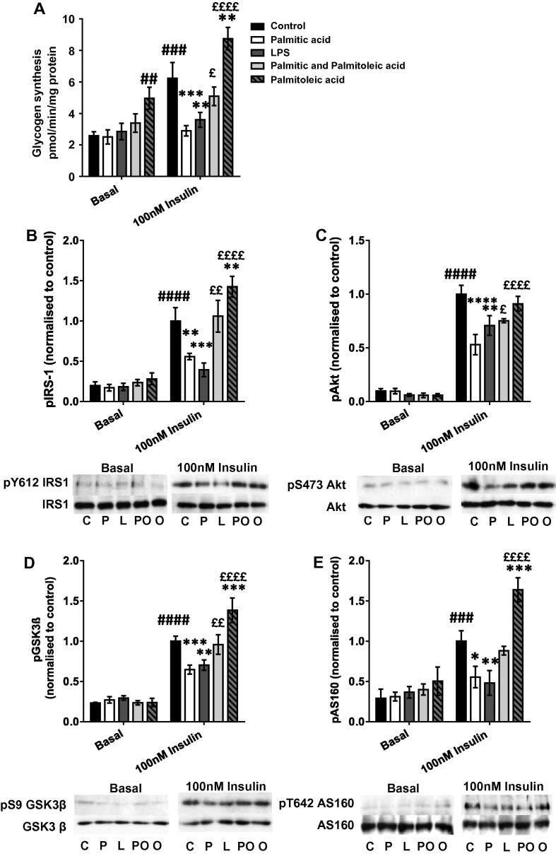
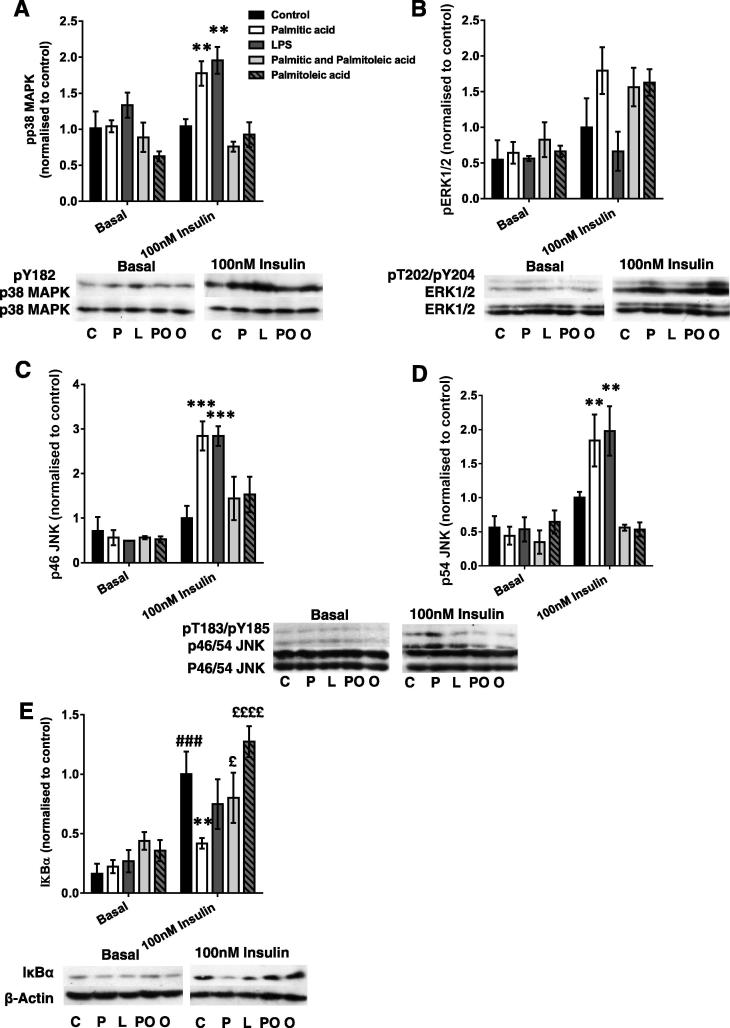
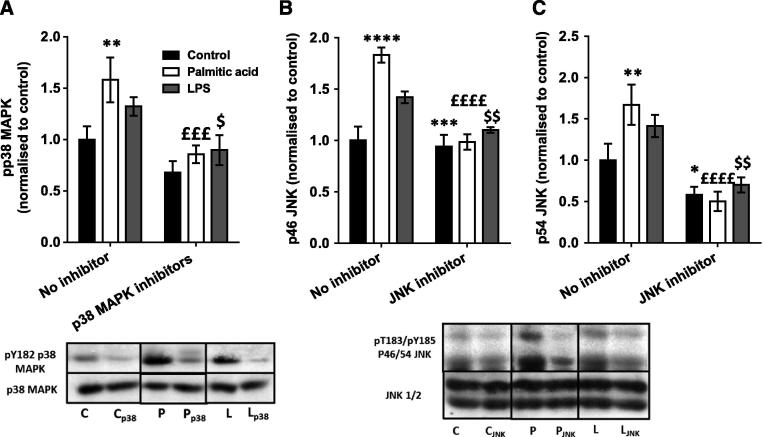
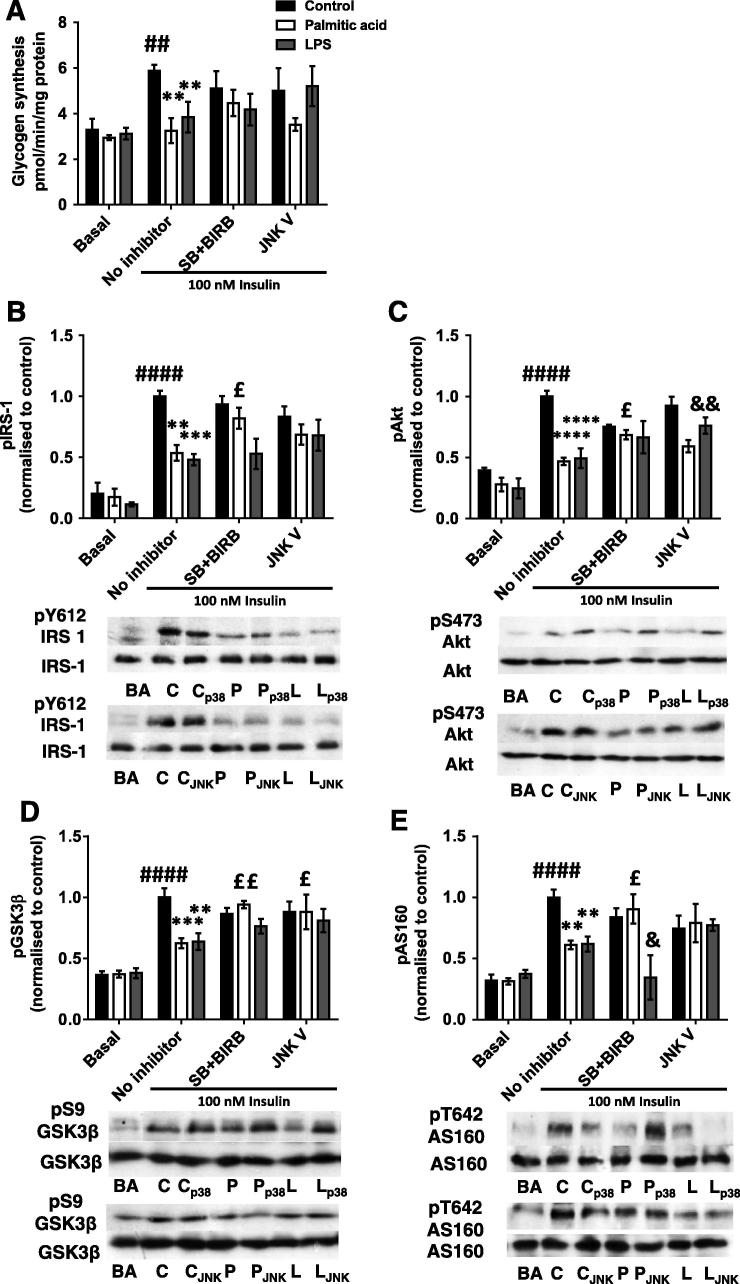
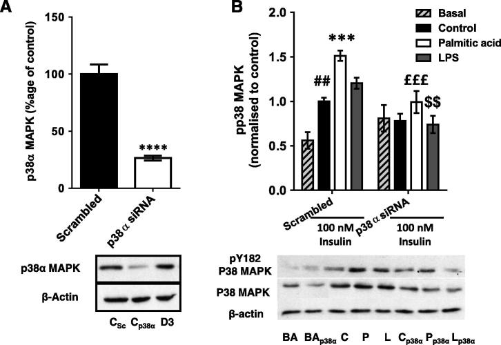
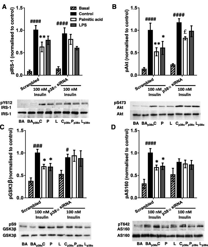
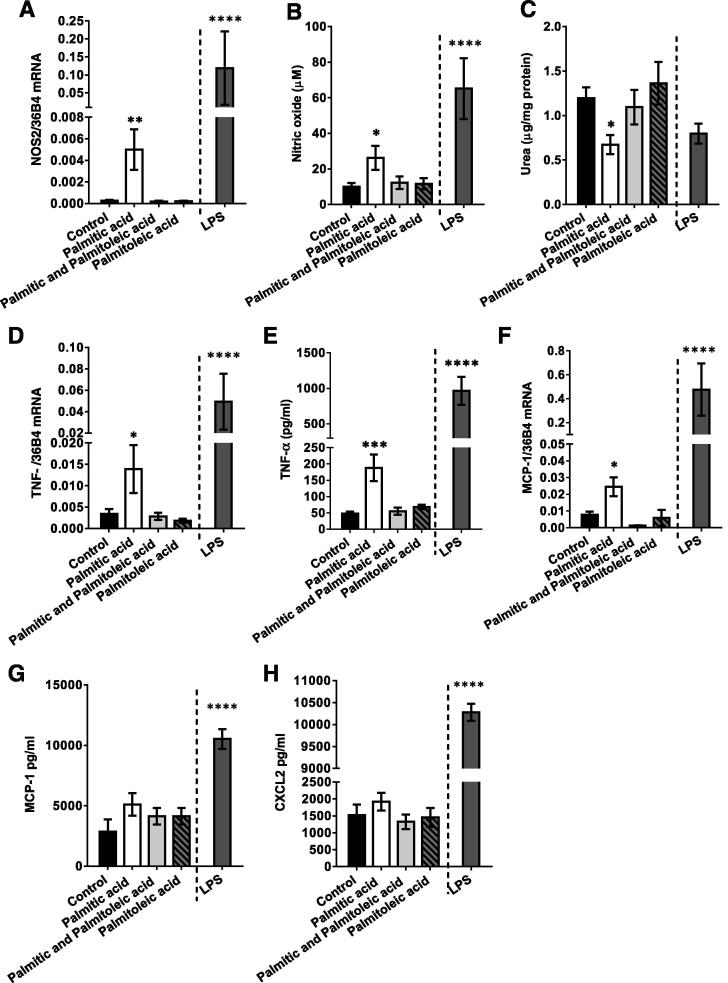
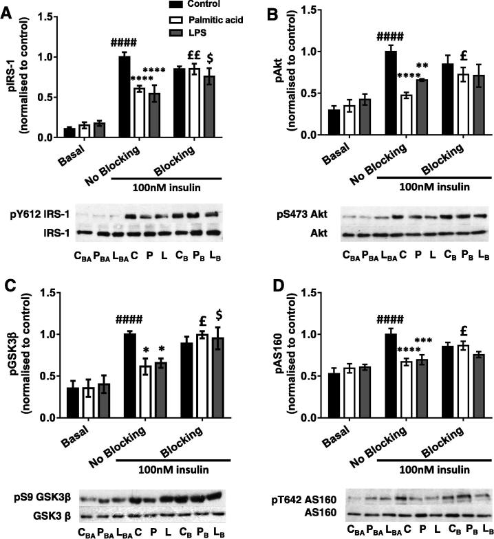
Obesity and saturated fatty acid (SFA) treatment are both associated with skeletal muscle insulin resistance (IR) and increased macrophage infiltration. However, the relative effects of SFA and unsaturated fatty acid (UFA)-activated macrophages on muscle are unknown. Here, macrophages were treated with palmitic acid, palmitoleic acid or both and the effects of the conditioned medium (CM) on C2C12 myotubes investigated. CM from palmitic acid-treated J774s (palm-mac-CM) impaired insulin signalling and insulin-stimulated glycogen synthesis, reduced Inhibitor κBα and increased phosphorylation of p38 mitogen-activated protein kinase (MAPK) and c-Jun N-terminal kinase in myotubes. p38 MAPK inhibition or siRNA partially ameliorated these defects, as did addition of tumour necrosis factor-α blocking antibody to the CM. Macrophages incubated with both FAs generated CM that did not induce IR, while palmitoleic acid-mac-CM alone was insulin sensitising. Thus UFAs may improve muscle insulin sensitivity and counteract SFA-mediated IR through an effect on macrophage activation.

Keywords: Fatty acid; Insulin resistance; Macrophage; Skeletal muscle; Tumour necrosis factor-α; p38 Mitogen-activated protein kinase.


Figures

Fig. 1
Fig. 1
Palmitic acid-treated macrophage-conditioned medium impairs glycogen synthesis and insulin signalling in C2C12 myotubes, while these defects are rescued by palmitoleic acid. C2C12 myotubes were incubated with conditioned medium derived from macrophages treated with LPS, palmitic acid, palmitoleic acid, a combination of the two or vehicle (control group) for 16 h, before being serum starved and (A) incubated with D-[U-14C]-glucose tracer ± 100nM insulin to measure glycogen synthesis, or collected in RIPA buffer and SDS–PAGE and immunoblotting conducted to assess phosphorylation and total protein levels of (B) IRS1 (pY612), (C) Akt (pS473), (D) GSK3β (pS9) and (E) AS160 (pT642). Representative blots and summary data (mean ± SEM of 3–6 individual experiments) are shown. All treatment groups were represented on each blot on which bands were quantified, but basal and insulin-stimulated samples are shown separately here for clarity. Total protein levels of all intermediates and β-actin were unchanged by any treatment. Selected post hoc significance is shown to simplify interpretation: ##p < 0.01, ###p > 0.001 and ####p < 0.0001 vs. basal control; *p < 0.05, **p < 0.01, ***p < 0.001 and ****p < 0.0001 vs. control, insulin treated cells; £p< 0.05, ££p < 0.01 and ££££p < 0.0001 versus palmitate treated, insulin treated cells. C – Control (+ insulin treatment); P – Palmitic acid (+ insulin treatment); L – LPS (+ insulin treatment); PO – Palmitic and palmitoleic acid (+ insulin treatment); O – Palmitoleic acid (+ insulin treatment).
Fig. 2
Fig. 2
Palmitic acid and palmitoleic acid-treated macrophage-conditioned media have contrasting effects on inflammatory signalling in C2C12 myotubes. C2C12 myotubes were incubated with conditioned medium derived from macrophages treated with LPS, palmitic acid, palmitoleic acid, a combination of the two or vehicle (control group) for 16 h, before being serum starved and lysed in RIPA buffer, and SDS–PAGE and immunoblotting conducted to assess phosphorylation and total protein levels of (A) p38 MAPK (pY182), (B) ERK1/2 (pT202), (C) p46 JNK (pT183/pY185), (D) p54 JNK (pT183/pY185) and E) IκBα. Densitometry was performed and sample blots and summary graphs are shown, representing the mean ± SEM of 3–4 individual experiments. All treatment groups were represented on each blot on which bands were quantified, but basal and insulin-stimulated samples are shown separately here for clarity. Total protein levels of MAPKs and β-Actin were unchanged by the treatments. Selected post hoc significance is shown to simplify interpretation: *p < 0.05, **p < 0.01, ***p < 0.001 vs. insulin-treated control; ###p < 0.001 vs. basal control; £p < 0.05, and ££££p < 0.0001 versus palmitate treated, insulin treated cells. C – Control (+ insulin treatment); P –Palmitic acid (+ insulin treatment); L – LPS (+ insulin treatment); PO – Palmitic and palmitoleic acid (+ insulin treatment); O – Palmitoleic acid (+ insulin treatment).
Fig. 3
Fig. 3
Effects of pharmacological inhibition of MAPKs on conditioned medium-induced changes in MAPK phosphorylation. Conditioned medium from macrophages treated with palmitic acid, LPS or vehicle (control group) was incubated with or without SB205380 (1 μM) and BIRB796 (0.1 μM), JNK V (1 μM) inhibitors or vehicle for 16 h. After cells were serum starved for 1 h and treated with 100nM insulin for 15 min, they were collected in RIPA buffer and phosphorylation and total protein levels of (A) p38 MAPK (pY182), (B) p46 JNK (pT183/pY185) and (C) p54 JNK (pT183/pY185) were assessed by SDS–PAGE and immunoblotting. Summary graphs show the mean ± SEM of 4 individual experiments, accompanied by representative blots. Total protein levels of signalling intermediates and β-Actin were unchanged by the treatments. Post hoc: *p < 0.05, **p < 0.01 and ***p < 0.001 vs. no inhibitor control; £££p < 0.001 vs. no inhibitor palmitic acid; $p < 0.05 and $$p < 0.01 vs. no inhibitor LPS. C – control conditioned medium (+ insulin treatment); P – palmitic acid-treated macrophage-conditioned medium (+ insulin treatment); L – LPS-treated macrophage-conditioned medium (+ insulin treatment); p38 – SB205380 and BIRB796 inhibitors used; JNK – JNK V inhibitor used.
Fig. 4
Fig. 4
Macrophage-conditioned medium-induced defects in glycogen synthesis and insulin signalling are partially restored by pharmacological MAPK inhibition. C2C12 myotubes were incubated with conditioned medium from macrophages treated with palmitic acid, LPS or control with or without SB205380 (1 μM) and BIRB796 (both 0.1 μM; “SB + BIRB”), JNK V (1 μM) inhibitors or vehicle (“No inhibitor” group) for 16 h, before being serum starved and (A) incubated with D-[U-14C]-glucose tracer ± 100nM insulin to measure glycogen synthesis, or collected in RIPA buffer and SDS–PAGE and immunoblotting conducted to assess phosphorylation and total protein levels of (B) IRS-1 (pY612), (C) Akt (pS473), (D) GSK3b (pS9) and (E) AS160 (pT642). Densitometry was performed and these data are shown graphically alongside representative blots. Total protein levels of signalling intermediates and β-Actin were unchanged by the treatments. Data are mean ± SEM of 4–6 individual experiments. Selected post hoc significance is shown to simplify interpretation: *p < 0.05, **p < 0.01 ***p < 0.001 and ****p < 0.0001 vs. no inhibitor insulin-stimulated control; £p < 0.05, ££p < 0.01 vs. no inhibitor palmitic acid; &p < 0.05 and &&p < 0.01 vs. no inhibitor LPS; ##p < 0.01 and ####p < 0.0001 vs. basal (no insulin) control. C – control conditioned medium (+insulin); P – palmitic acid-treated macrophage-conditioned medium (+ insulin): L – LPS-treated macrophage-conditioned medium (+ insulin). BA – basal (no insulin); p38 – SB205380 and BIRB796 inhibitors used; JNK – JNK V inhibitor used.
Fig. 5
Fig. 5
Effects of siRNA-mediated p38 MAPKα silencing on conditioned medium-induced changes in p38 MAPK phosphorylation and protein levels in C2C12 myotubes. Western blots were generated using lysates derived from myotubes incubated with a siRNA pool targeting p38 MAPKα or a nonsense (scrambled) control for 72 h. (A) Silencing of p38α MAPK protein in 100nM insulin-treated C2C12 myotubes (mean 74% reduction). (B) C2C12 myotubes were incubated with conditioned medium generated by macrophages treated with palmitic acid, LPS or vehicle (control group) ± p38α siRNA pool for 16 h. Cells were serum starved for 1 h and then treated ± 100nM insulin for 15 min. Total phosphorylation and total protein levels of p38 MAPK (all isoforms) were assessed. Summary graphs show the mean ± SEM of 4 individual experiments, accompanied by representative blots. Selected post hoc significance is shown to simplify interpretation: ##p < 0.01 vs. scrambled siRNA, basal ***p < 0.001 and ****p < 0.0001 vs. scrambled siRNA, insulin-treated; £££p < 0.001 vs. scrambled siRNA palmitic acid; $$p < 0.01 vs. scrambled siRNA LPS. C – control conditioned medium (+ insulin treatment); P – palmitic acid-treated macrophage-conditioned medium (+ insulin treatment); L – LPS-treated macrophage-conditioned medium (+ insulin treatment). BA – basal (no insulin); p38α – p38 MAPKα siRNA-treated; D3 – transfection reagent-treated only.
Fig. 6
Fig. 6
The palmitic acid-treated macrophage-CM-induced defect in myotube PI3K signalling is partially restored by siRNA-induced silencing of p38α. Conditioned medium from macrophages treated with palmitic acid, LPS or vehicle (control group) was used to incubate C2C12 myotubes ± a p38 MAPKα siRNA pool for 16 h. Cells were serum starved for 1 h and treated ± 100nM insulin for 15 min. Phosphorylation and protein levels of (A) IRS-1 (pY612), (B) Akt (pS473), (C) GSK3β (pS9) and (D) AS160 (pT642) were assessed by western blotting of lysates. Densitometry was performed on blots and summarised as graphs showing the mean ± SEM of 4 individual experiments, alongside representative blots. Total protein levels were unchanged by any treatment. Selected post hoc significance is shown to simplify interpretation: *p < 0.05 and **p < 0.01 vs. scrambled siRNA insulin-treated control; £p < 0.05 vs. scrambled siRNA palmitic acid; #p < 0.05, ###p < 0.001 and ####p < 0.0001 vs. basal (no insulin) sample from the same siRNA treatment group. C – control conditioned medium (+ insulin); P – palmitic acid-treated macrophage-conditioned medium (+ insulin); L – LPS-treated macrophage-conditioned medium (+ insulin). BA – basal (no insulin); p38α – p38 MAPKα siRNA-treated.
Fig. 7
Fig. 7
Macrophages treated with palmitic acid show a pro-inflammatory polarisation that is abrogated by the addition of palmitoleic acid. J774 Macrophages were incubated with 0.75 mM palmitic acid, 0.75 mM palmitoleic acid, a combination of both, LPS, or vehicle (control group) for 8 h, washed with PBS and then conditioned medium (CM) generated for 16 h. After this period, CM and macrophages were collected and (A) macrophage mRNA expression of NOS2 was measured, (B) content of nitric oxide (μM) in culture medium was measured using the Griess assay, (C) arginase activity was assessed based on the amount of urea (μg) produced per mg of protein, (D) TNFα mRNA and (E) TNFα peptide were measured in macrophages/CM respectively by real-time PCR/ELISA, (F) MCP1 mRNA and G) MCP1 peptide were measured in macrophages/CM respectively and (H) CXCL2 peptide was measured in CM by ELISA. Values are the mean ± SEM of 5–6 individual experiments. Post hoc: *p < 0.05, ***p < 0.001 and ****p < 0.0001 vs. control.
Fig. 8
Fig. 8
Addition of a TNFα blocking antibody leads to partial restoration of the palmitic acid-treated macrophage CM-induced defect in myotube insulin signalling. C2C12 myotubes were incubated with conditioned medium derived from macrophages treated with palmitic acid, LPS or vehicle (No blocking group), with or without TNFα blocking antibody (10 μg/ml) for 16 h, before being serum starved and lysed in RIPA buffer, and SDS–PAGE and immunoblotting conducted to assess phosphorylation and total protein levels of (A) IRS1 (pY612), (B) Akt (pS473), (C) GSK3β (pS9) and (D) AS160 (pT642). Representative blots and summary data (mean ± SEM of 6 individual experiments) are shown. Protein expression levels of all intermediates were unchanged by any treatment. Selected post hoc significance is shown to simplify interpretation: *p < 0.05, **p < 0.01, ***p < 0.001 ****p < 0.0001 vs. insulin-treated control; £p < 0.05; ££p < 0.01 vs. No Blocking palmitic acid; $p < 0.05 vs No Blocking LPS; ####p < 0.0001 vs. Basal control. B – Basal (no insulin) C – Control, insulin-treated P – Palmitic acid, insulin treated, L – LPS, insulin treated. BA – Basal, B – Blocking antibody present.

Similar articles

Cited by

References

    1. Aguirre V., Werner E.D., Giraud J., Lee Y.H., Shoelson S.E., White M.F. Phosphorylation of Ser307 in insulin receptor substrate-1 blocks interactions with the insulin receptor and inhibits insulin action. J. Biol. Chem. 2002;277:1531–1537. doi: 10.1074/jbc.M101521200. - DOI - PubMed
    1. Antonescu C.N., Huang C., Niu W., Liu Z., Eyers P.A., Heidenreich K.A., Bilan P.J., Klip A. Reduction of insulin-stimulated glucose uptake in L6 myotubes by the protein kinase inhibitor SB203580 is independent of p38MAPK activity. Endocrinology. 2005;146:3773–3781. doi: 10.1210/en.2005-0404. - DOI - PubMed
    1. Bain J., Plater L., Elliott M., Shpiro N., Hastie C.J., McLauchlan H., Klevernic I., Arthur J.S., Alessi D.R., Cohen P. The selectivity of protein kinase inhibitors: a further update. Biochem. J. 2007;408:297–315. doi: 10.1042/BJ20070797. - DOI - PMC - PubMed
    1. Boppart M.D., Asp S., Wojtaszewski J.F., Fielding R.A., Mohr T., Goodyear L.J. Marathon running transiently increases c-Jun NH2-terminal kinase and p38 activities in human skeletal muscle. J. Physiol. 2000;526(Pt 3):663–669. doi: 10.1111/j.1469-7793.2000.00663.x. - DOI - PMC - PubMed
    1. Cancello R., Henegar C., Viguerie N., Taleb S., Poitou C., Rouault C., Coupaye M., Pelloux V., Hugol D., Bouillot J.L., Bouloumie A., Barbatelli G., Cinti S., Svensson P.A., Barsh G.S., Zucker J.D., Basdevant A., Langin D., Clement K. Reduction of macrophage infiltration and chemoattractant gene expression changes in white adipose tissue of morbidly obese subjects after surgery-induced weight loss. Diabetes. 2005;54:2277–2286. doi: 10.2337/diabetes.54.8.2277. - DOI - PubMed

Publication types

MeSH terms