

Percutaneous closure of complex paravalvular aortic root pseudoaneurysm and aorta-cavitary fistulas
- PMID: 24973845
- PMCID: PMC4121747
- DOI: 10.1016/j.ihj.2014.03.015
Percutaneous closure of complex paravalvular aortic root pseudoaneurysm and aorta-cavitary fistulas
Abstract
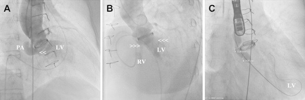
Native aortic valve or its prosthetic valve endocarditis can extend to the adjacent periannular areas and erode into nearby cardiac chambers, leading to pseudoaneurysm and aorta-cavitary fistulas respectively. The later usually leads to acute cardiac failure and hemodynamic instability requiring an urgent surgical intervention. However rarely this might pass unnoticed and the patient might present later with cardiac murmur. Percutaneous device closure of aortic pseudoaneurysm, ruptured sinus of Valsalva aneurysm, aorta-pulmonary window, paravalvular leaks, and aorta-cavitary fistula have been reported. We present a 59-year-old female who developed a large aortic root pseudoaneurysm with biventricular communication aorta-cavitary fistulas presenting late following aortic prosthetic valve endocarditis. She underwent successful percutaneous device closure of her pseudoaneurysm and aorta-cavitary fistulas using two Amplatzer Duct Occluders. This case illustrates a challenging combination of aortic root pseudoaneurysm and biventricular aorta-cavitary fistulas that was successfully treated with percutaneous procedure.
Keywords: Aorta-cavitary fistula; Device closure; Endocarditis; Pseudoaneurysm.
Copyright © 2014 Cardiological Society of India. Published by Elsevier B.V. All rights reserved.
Figures


Similar articles
-
Percutaneous repair of ascending aorta pseudoaneurysm and aortopulmonary fistula with two Amplatzer septal occluder devices.Eur Heart J. 2017 Oct 1;38(37):2853-2854. doi: 10.1093/eurheartj/ehw579. Eur Heart J. 2017. PMID: 28025187 No abstract available.
-
Transcatheter Closure of Ruptured Sinus of Valsalva Aneurysm with Double-Disc Perimembranous VSD Occluder in Man with Mechanical Aortic Valve.Tex Heart Inst J. 2019 Jun 1;46(3):211-214. doi: 10.14503/THIJ-17-6215. eCollection 2019 Jun. Tex Heart Inst J. 2019. PMID: 31708706 Free PMC article.
-
Comparison of immediate results and mid-term follow-up of surgical and percutaneous closure of ruptured sinus of Valsalva aneurysm.J Cardiol. 2014 Mar;63(3):239-43. doi: 10.1016/j.jjcc.2013.08.011. Epub 2013 Oct 20. J Cardiol. 2014. PMID: 24148861
-
Endovascular exclusion of complex postsurgical aortic arch pseudoaneurysm using vascular plug devices and a review of vascular plugs.Perspect Vasc Surg Endovasc Ther. 2012 Dec;24(4):193-7. doi: 10.1177/1531003513501203. Perspect Vasc Surg Endovasc Ther. 2012. PMID: 24052323 Review.
-
Use of Amplatzer occluders for treatment of aorto-pulmonary fistulas - case and review of the literature.Expert Rev Med Devices. 2017 Nov;14(11):845-847. doi: 10.1080/17434440.2017.1389636. Epub 2017 Oct 12. Expert Rev Med Devices. 2017. PMID: 29022410 Review.
Cited by
-
Endovascular Closure of 2 Subannular Pseudoaneurysms of the Aortic Root After Surgical Aortic Valve Replacement.JACC Case Rep. 2019 Dec 18;1(5):807-810. doi: 10.1016/j.jaccas.2019.11.011. eCollection 2019 Dec 18. JACC Case Rep. 2019. PMID: 34316936 Free PMC article.
-
Transesophageal echocardiographic guidance for percutaneous closure of aortic pseudoaneurysm using a type II Amplatzer vascular plug: a case report.Korean J Anesthesiol. 2016 Aug;69(4):400-5. doi: 10.4097/kjae.2016.69.4.400. Epub 2016 Jul 1. Korean J Anesthesiol. 2016. PMID: 27482320 Free PMC article.
-
Percutaneous device closure of a large complex aortic root pseudoaneurysm.BMJ Case Rep. 2020 Sep 17;13(9):e235545. doi: 10.1136/bcr-2020-235545. BMJ Case Rep. 2020. PMID: 32943443 Free PMC article.
References
-
- Kanemitsu S., Tanabe S., Ohue K., Miyagawa H., Miyake Y., Okabe M. Aortic valve destruction and pseudoaneurysm of the sinus of valsalva associated with infective endocarditis. Ann Thorac Cardiovasc Surg. 2010;16:142–144. - PubMed
-
- Anguera I., Miro J.M., Vilacosta I. Aorto-cavitary fistulous tract formation in infective endocarditis: clinical and echocardiographic features of 76 cases and risk factors for mortality. Eur Heart J. 2005;26:288–297. - PubMed
-
- Katayama Y., Minato N., Sakaguchi M., Nakashima A., Hisajima K. Surgical treatment of pseudoaneurysm of the sinus of valsalva after aortic valve replacement for active infective endocarditis. Ann Thorac Cardiovasc Surg. 2005;11:419–423. - PubMed
-
- Hori D., Noguchi K., Nomura Y., Tanaka H. Perivalvular pseudoaneurysm caused by streptococcus dysgalactiae in the presence of prosthetic aortic valve endocarditis. Ann Thorac Cardiovasc Surg. 2012;18:262–265. - PubMed
Publication types
MeSH terms
LinkOut - more resources
Full Text Sources
Other Literature Sources
Medical
