

Early results of a novel technique for anterior vaginal wall prolapse repair: anterior vaginal wall darn
- PMID: 24973955
- PMCID: PMC4105512
- DOI: 10.1186/1471-2490-14-51
Early results of a novel technique for anterior vaginal wall prolapse repair: anterior vaginal wall darn
Abstract
Background: The aim of this study was to describe the results of a 1-year patient follow-up after anterior vaginal wall darn, a novel technique for the repair of anterior vaginal wall prolapse.
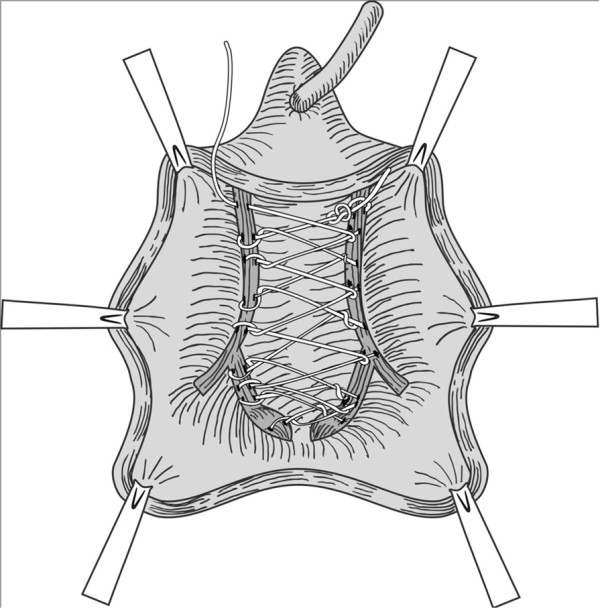
Methods: Fifty-five patients with anterior vaginal wall prolapse underwent anterior vaginal wall darn. The anterior vaginal wall was detached using sharp and blunt dissection via an incision beginning 1 cm proximal to the external meatus and extending to the vaginal apex. The space between the tissues that attach the lateral vaginal walls to the arcus tendineus fasciae pelvis was then darned. Cough Stress Test, Pelvic Organ Prolapse Quantification, seven-item Incontinence Impact Questionnaire, and six-item Urogenital Distress Inventory scores were performed 1-year postoperatively to evaluate recovery.
Results: One-year postoperatively, all patients were satisfied with the results of the procedure. No patient had vaginal mucosal erosion or any other complication.
Conclusions: One-year postoperative findings for patients in this series indicate that patients with stage II-III anterior vaginal wall prolapse were successfully treated with the anterior vaginal wall darn technique.
Figures


References
-
- Kenton K, Mueller ER. The global burden of female pelvic floor disorders. BJU Int. 2006;98:1–5. - PubMed
-
- Melville JL, Fan M, Rau H, Nygaard IE, Katon WJ. Major depression and urinary incontinence in women: temporal associations in an epidemiologic sample. Am J Obstet Gynecol. 2009;201(490):e1–e7. - PubMed
MeSH terms
LinkOut - more resources
Full Text Sources
Other Literature Sources
Medical
Miscellaneous
