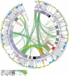
Distribution of segmental duplications in the context of higher order chromatin organisation of human chromosome 7
- PMID: 24973960
- PMCID: PMC4092221
- DOI: 10.1186/1471-2164-15-537
Distribution of segmental duplications in the context of higher order chromatin organisation of human chromosome 7
Abstract
Background: Segmental duplications (SDs) are not evenly distributed along chromosomes. The reasons for this biased susceptibility to SD insertion are poorly understood. Accumulation of SDs is associated with increased genomic instability, which can lead to structural variants and genomic disorders such as the Williams-Beuren syndrome. Despite these adverse effects, SDs have become fixed in the human genome. Focusing on chromosome 7, which is particularly rich in interstitial SDs, we have investigated the distribution of SDs in the context of evolution and the three dimensional organisation of the chromosome in order to gain insights into the mutual relationship of SDs and chromatin topology.
Results: Intrachromosomal SDs preferentially accumulate in those segments of chromosome 7 that are homologous to marmoset chromosome 2. Although this formerly compact segment has been re-distributed to three different sites during primate evolution, we can show by means of public data on long distance chromatin interactions that these three intervals, and consequently the paralogous SDs mapping to them, have retained their spatial proximity in the nucleus. Focusing on SD clusters implicated in the aetiology of the Williams-Beuren syndrome locus we demonstrate by cross-species comparison that these SDs have inserted at the borders of a topological domain and that they flank regions with distinct DNA conformation.
Conclusions: Our study suggests a link of nuclear architecture and the propagation of SDs across chromosome 7, either by promoting regional SD insertion or by contributing to the establishment of higher order chromatin organisation themselves. The latter could compensate for the high risk of structural rearrangements and thus may have contributed to their evolutionary fixation in the human genome.
Figures


Similar articles
-
Genomic regions associated with microdeletion/microduplication syndromes exhibit extreme diversity of structural variation.Genetics. 2021 Feb 9;217(2):iyaa038. doi: 10.1093/genetics/iyaa038. Genetics. 2021. PMID: 33724415 Free PMC article.
-
Evolutionary mechanisms shaping the genomic structure of the Williams-Beuren syndrome chromosomal region at human 7q11.23.Genome Res. 2005 Sep;15(9):1179-88. doi: 10.1101/gr.3944605. Genome Res. 2005. PMID: 16140988 Free PMC article.
-
Fine-scale comparative mapping of the human 7q11.23 region and the orthologous region on mouse chromosome 5G: the low-copy repeats that flank the Williams-Beuren syndrome deletion arose at breakpoint sites of an evolutionary inversion(s).Genomics. 2000 Oct 1;69(1):1-13. doi: 10.1006/geno.2000.6312. Genomics. 2000. PMID: 11013070
-
The genomic basis of the Williams-Beuren syndrome.Cell Mol Life Sci. 2009 Apr;66(7):1178-97. doi: 10.1007/s00018-008-8401-y. Cell Mol Life Sci. 2009. PMID: 19039520 Free PMC article. Review.
-
Williams-Beuren syndrome: genes and mechanisms.Hum Mol Genet. 1999;8(10):1947-54. doi: 10.1093/hmg/8.10.1947. Hum Mol Genet. 1999. PMID: 10469848 Review.
Cited by
-
Intrauterine phenotype features of fetuses with 7q11.23 microduplication syndrome.Orphanet J Rare Dis. 2023 Sep 27;18(1):305. doi: 10.1186/s13023-023-02923-y. Orphanet J Rare Dis. 2023. PMID: 37759207 Free PMC article.
-
A Polymer Physics Investigation of the Architecture of the Murine Orthologue of the 7q11.23 Human Locus.Front Neurosci. 2017 Oct 10;11:559. doi: 10.3389/fnins.2017.00559. eCollection 2017. Front Neurosci. 2017. PMID: 29066944 Free PMC article. Review.
-
5'RUNX1-3'USP42 chimeric gene in acute myeloid leukemia can occur through an insertion mechanism rather than translocation and may be mediated by genomic segmental duplications.Mol Cytogenet. 2014 Oct 1;7(1):66. doi: 10.1186/s13039-014-0066-7. eCollection 2014. Mol Cytogenet. 2014. PMID: 25298786 Free PMC article.
-
Distal 7q11.23 Duplication, an Emerging Microduplication Syndrome: A Case Report and Further Characterisation.Mol Syndromol. 2016 Oct;7(5):287-291. doi: 10.1159/000448698. Epub 2016 Aug 24. Mol Syndromol. 2016. PMID: 27867344 Free PMC article.
References
-
- Marques-Bonet T, Kidd JM, Ventura M, Graves TA, Cheng Z, Hillier LW, Jiang Z, Baker C, Malfavon-Borja R, Fulton LA, Alkan C, Aksay G, Girirajan S, Siswara P, Chen L, Cardone MF, Navarro A, Mardis ER, Wilson RK, Eichler EE. A burst of segmental duplications in the genome of the African great ape ancestor. Nature. 2009;457:877–881. doi: 10.1038/nature07744. - DOI - PMC - PubMed
-
- She X, Liu G, Ventura M, Zhao S, Misceo D, Roberto R, Cardone MF, Rocchi M, Program NCS, Green ED, Archidiacano N, Eichler EE. A preliminary comparative analysis of primate segmental duplications shows elevated substitution rates and a great-ape expansion of intrachromosomal duplications. Genome Res. 2006;16:576–583. doi: 10.1101/gr.4949406. - DOI - PMC - PubMed
Publication types
MeSH terms
Substances
Supplementary concepts
Associated data
- Actions
- SRA/SRS366467
LinkOut - more resources
Full Text Sources
Other Literature Sources
Molecular Biology Databases

