Determining absolute protein numbers by quantitative fluorescence microscopy
- PMID: 24974037
- PMCID: PMC4221264
- DOI: 10.1016/B978-0-12-420138-5.00019-7
Determining absolute protein numbers by quantitative fluorescence microscopy
Abstract
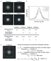
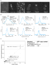
Biological questions are increasingly being addressed using a wide range of quantitative analytical tools to examine protein complex composition. Knowledge of the absolute number of proteins present provides insights into organization, function, and maintenance and is used in mathematical modeling of complex cellular dynamics. In this chapter, we outline and describe three microscopy-based methods for determining absolute protein numbers--fluorescence correlation spectroscopy, stepwise photobleaching, and ratiometric comparison of fluorescence intensity to known standards. In addition, we discuss the various fluorescently labeled proteins that have been used as standards for both stepwise photobleaching and ratiometric comparison analysis. A detailed procedure for determining absolute protein number by ratiometric comparison is outlined in the second half of this chapter. Counting proteins by quantitative microscopy is a relatively simple yet very powerful analytical tool that will increase our understanding of protein complex composition.
Keywords: Counting; FCS; Fluorescence; Fluorescence standards; Photobleaching; Quantitative imaging; Ratiometric.
© 2014 Elsevier Inc. All rights reserved.
Figures


References
-
- Aravamudhan P, Felzer-Kim I, Joglekar AP. The budding yeast point centromere associates with two Cse4 molecules during mitosis. Current Biology. 2013;23(9):770–774. http://dx.doi.org/10.1016/j.cub.2013.03.042. - DOI - PMC - PubMed
-
- Bacia K, Schwille P. A dynamic view of cellular processes by in vivo fluorescence auto- and cross-correlation spectroscopy. Methods. 2003;29(1):74–85. - PubMed
-
- Braeckmans K, Deschout H, Demeester J, De Smedt SC. Optical fluorescence microscopy: From the spectral to the nano dimension. Berlin, Heidelberg: Springer-Verlag; 2011. Measuring molecular dynamics by FRAP, FCS, and SPT; pp. 153–163.
-
- Bulseco DA, Wolf DE. Fluorescence correlation spectroscopy: Molecular complexing in solution and in living cells. Methods in Cell Biology. 2013;114:489–524. http://dx.doi.org/10.1016/B978-0-12-407761-4.00021-X. - DOI - PubMed
-
- Charpilienne A, Nejmeddine M, Berois M, Parez N, Neumann E, Hewat E, et al. Individual rotavirus-like particles containing 120 molecules of fluorescent protein are visible in living cells. The Journal of Biological Chemistry. 2001;276(31):29361–29367. http://dx.doi.org/10.1074/jbc.M101935200. - DOI - PubMed
MeSH terms
Substances
Grants and funding
LinkOut - more resources
Full Text Sources
Other Literature Sources

