. 2014 Jun 29;15(1):538.
doi: 10.1186/1471-2164-15-538.

Transcriptome dynamics of Arabidopsis thaliana root penetration by the oomycete pathogen Phytophthora parasitica

Affiliations

Transcriptome dynamics of Arabidopsis thaliana root penetration by the oomycete pathogen Phytophthora parasitica

Agnès Attard et al. BMC Genomics. .

Abstract

Background: Oomycetes are a group of filamentous microorganisms that includes both animal and plant pathogens and causes major agricultural losses. Phytophthora species can infect most crops and plants from natural ecosystems. Despite their tremendous economic and ecologic importance, few effective methods exist for limiting the damage caused by these species. New solutions are required, and their development will require improvements in our understanding of the molecular events governing infection by these pathogens. In this study, we characterized the genetic program activated during penetration of the plant by the soil-borne pathogen Phytophthora parasitica.

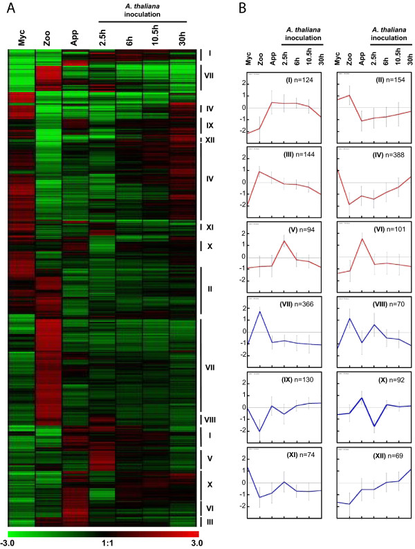
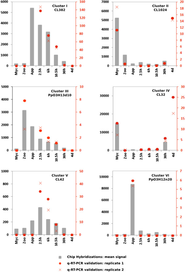
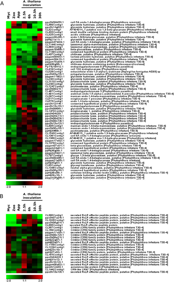
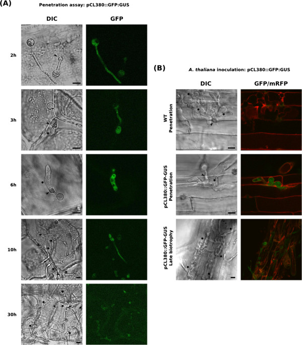
Results: Using all the P. parasitica sequences available in public databases, we generated a custom oligo-array and performed a transcriptomic analysis of the early events of Arabidopsis thaliana infection. We characterized biological stages, ranging from the appressorium-mediated penetration of the pathogen into the roots to the occurrence of first dead cells in the plant. We identified a series of sequences that were transiently modulated during host penetration. Surprisingly, we observed an overall down regulation of genes encoding proteins involved in lipid and sugar metabolism, and an upregulation of functions controlling the transport of amino acids. We also showed that different groups of genes were expressed by P. parasitica during host penetration and the subsequent necrotrophic phase. Differential expression patterns were particularly marked for cell wall-degrading enzymes and other proteins involved in pathogenicity, including RXLR effectors. By transforming P. parasitica with a transcriptional fusion with GFP, we showed that an RXLR-ecoding gene was expressed in the appressorium and infectious hyphae during infection of the first plant cell.

Conclusion: We have characterized the genetic program activated during the initial invasion of plant cells by P. parasitica. We showed that a specific set of proteins, including effectors, was mobilized for penetration and to facilitate infection. Our detection of the expression of an RXLR encoding gene by the appressorium and infection hyphae highlights a role of this structure in the manipulation of the host cells.


Figures

Figure 1
Figure 1
Hierarchical and K-mean clustering of four-fold expressed genes. (A) Hierarchical clustering of the 1806 genes modulated by 4-fold. Pearson correlation method with average linkage on conditions was used. Gene expression level is indicated as a Log2 transformation of relative value calculated on gene average signal value. Green and red were used for down-regulated and up-regulated conditions respectively. Lines mark major K-mean groups. (B) Expression profiles were K-mean clustered based on euclidean distance. The twelve main K-mean clusters are represented. The center line represents average expression with standard deviation. Number of genes within each cluster is indicated. The six clusters used for subsequent characterization of the molecular events occurring during early infection are colored in red. Myc, mycelium; Zoo, zoospores, App, Appressorium; 2.5 h, Arabidopsis thaliana infected roots recovered 2.5 hours after inoculation (appressorium-mediated penetration); 6 h, A. thaliana infected roots recovered 6 hours after inoculation (biotrophic growth, two to three cells invaded); 10.5 h, A. thaliana infected roots recovered 10.5 hours after inoculation (invasive growth along the stele); 30 h, A. thaliana infected roots recovered 30 hours after inoculation (switch to necrotrophy).
Figure 2
Figure 2
Quantification of mRNA corresponding to on gene of each of the clusters I to VI. Relative mRNA levels were quantified by quantitative RT-PCR in biological replicates of samples used in Figure 1. A condition corresponding to A. thaliana roots 4 days after inoculation (4d), corresponding to necrotrophic stage of the interaction was added. Data are presented as expression ratios relative to UBC, WS21 and Mago Nashi reference genes (2−ΔCT). Grey bars represent mean signal obtained following oligoarray hybridizations (black left axis). Red point and crosses represent relative expression values obtained for two independent biological replicates analyzed using qRT-PCR (red right axis).
Figure 3
Figure 3
Hierarchical clustering of the genes encoding cell wall degrading enzymes and cytoplasmic effectors. Based on GO annotations, sequences corresponding to cell wall degrading enzymes (A) and sequences corresponding to RXLR and Crinkler effectors (B) are presented. Pearson correlation method with average linkage on conditions was used. Gene expression level is indicated as a Log2 transformation of relative value calculated on gene average signal value. Green and red were used for down-regulated and up-regulated conditions respectively. Myc, mycelium; Zoo, zoospores, App, Appressorium; 2.5 h, A. thaliana infected roots recovered 2.5 hours after inoculation; 6 h, A. thaliana infected roots recovered 6 hours after inoculation; 10.5 h, A. thaliana infected roots recovered 10.5 hours after inoculation; 30 h, A. thaliana infected roots recovered 30 hours after inoculation.
Figure 4
Figure 4
P. Parasitica appressorium transiently express a RXLR encoding effector during the penetration process. (A and B) Expression pattern of the CL380 RXLR encoding gene using a pCL380::GFP-GUS fusion. (A) Kinetic of early infection steps using a simplified penetration assay based on onion epidermis. GFP fluorescence from a P. parasitica strain carrying the pCL380::GFP-GUS construct was monitored 2 to 30 hours after inoculation of zoospores on onion epidermis. (B) Analysis of the CL380 expression pattern during early A. thaliana infection. A. thaliana plantlets where inoculated with zoospores from a P. parasitica strain carrying the pCL380::GFP-GUS construct and fluorescence was monitored during infection of the first cell (6 hours after inoculation for the presented image) and late biotrophic phase (24 hours after inoculation for the proposed image). Plant cell membranes (red) are visualized with the mCherry plasma membrane marker pm-RB. A representative image for each stage is presented. GFP and mCherry fluorescence was visualized using a confocal laser scanning microscope. Bars: 10 μM; Arrows: appressoria; Stars: Infectious hyphae.

Similar articles

Cited by

References

    1. Erwin DC, Ribeiro OK. Phytophthora Diseases Worldwide. St. Paul, MN: American Phytopathological Society; 1996.
    1. Beakes GW, Glockling SL, Sekimoto S. The evolutionary phylogeny of the oomycete “fungi”. Protoplasma. 2011;249:3–19. doi: 10.1007/s00709-011-0269-2. - DOI - PubMed
    1. Cavalier-Smith T, Chao EE-Y. Phylogeny and Megasystematics of Phagotrophic Heterokonts (Kingdom Chromista) J Mol Evol. 2006;62:388–420. doi: 10.1007/s00239-004-0353-8. - DOI - PubMed
    1. Swiecki TJ, Donald M. Histology of chrysanthemum roots exposed to salinity stress and Phytophthora cryptogea. Can J Bot. 1988;66:280–288. doi: 10.1139/b88-046. - DOI
    1. Dale ML, Irwin JAG. Stomata as an infection court for Phytophtora megasperma f. sp. medicaginis in chickpea and a histological study of infection. Phytopathology. 1991;91:375–379. doi: 10.1094/Phyto-81-375. - DOI

Publication types

LinkOut - more resources