Review
. 2014 Aug;35(8):379-86.
doi: 10.1016/j.it.2014.05.005. Epub 2014 Jun 26.

The spatiotemporal cellular dynamics of lung immunity

Affiliations
Review

The spatiotemporal cellular dynamics of lung immunity

Efrat Lelkes et al. Trends Immunol. 2014 Aug.

Abstract

The lung is a complex structure that is interdigitated with immune cells. Understanding the 4D process of normal and defective lung function and immunity has been a centuries-old problem. Challenges intrinsic to the lung have limited adequate microscopic evaluation of its cellular dynamics in real time, until recently. Because of emerging technologies, we now recognize alveolar-to-airway transport of inhaled antigen. We understand the nature of neutrophil entry during lung injury and are learning more about cellular interactions during inflammatory states. Insights are also accumulating in lung development and the metastatic niche of the lung. Here we assess the developing technology of lung imaging, its merits for studies of pathophysiology and areas where further advances are needed.


Figures

Figure 1
Figure 1
Evolution of the thoracic window. (a) The original thoracic window as described by Terry in 1939 for use in cats. (From Terry, R. J. (1939) A Thoracic Window for Observation of the Lung in a Living Animal. Science. 90, 43–44. Reprinted with permission from AAAS.) (b) The thoracic window as described by Looney et al in 2011. Shown in panel is (i) the thoracic window itself, (ii) a schematic side-view representation of the vacuum suction and contact to coverslip and lungs, (iii) the surgical preparation and location of thoracic window on a mouse in the right lateral decubitus position with a left thoracotomy and an exposed left lung, and finally (iv) the surgical preparation of a mechanically ventilated mouse with thoracic window in place. (Reprinted by permission from Macmillan Publishers Ltd: Nature Methods. Looney, M.R. et al. Stabilized imaging of immune surveillance in the mouse lung.8(1) copyright 2011)
Figure 2
Figure 2. Challenges of imaging air-filled lungs
(a) The unobstructed view of a single alveolus in which rays from the microscope converge at a single point. (b) With a second alveolus and thus a second air-water interface, ideally rays would still converge at a single point. (c) In reality, with a second alveolus, rays interfere with each other and converge at distinct points. (d) Intravital imaging through a thoracic window in a mouse injected i.v. with Texas Red Dextran showing loss of resolution and signal at progressively deeper layers (22 um, 42 um, 90 um, and (bottom) showing a z-projection).
Figure 3
Figure 3. The immune system of the lung
(a) Epithelial cells line the airways to defend the host from antigen. Cilia mechanically move antigen out of the airway. Goblet cells produce mucous to help eliminate antigen. (b) The epithelium forms both a physical barrier to antigen entry into the lung and an immune barrier by its ability to incite an immune response to antigen. (c) The alveolus is a site of immune surveillance by alveolar macrophages within the alveolus, and by dendritic cells within the interstitum which extend processes into the alveolus. (d) Capillaries course in close proximity to alveoli composed of thin type I and type II alveolar epithelial cells and contain leukocytes, particularly neutrophils (in addition to erythrocytes, not illustrated) which marginate in the lungs searching for breach of immunity.
Figure 4
Figure 4. Lessons learned from lung imaging
(A) Alveolar dendritic cells send processes into alveoli where they take up antigen, seen both in schematic and in a 2-photon image of a lung slice in a CD11c-eYFP mouse, with dendritic cells in green (with arrow) (i) and then traffic up along airways where they interact with T cells in allergic airways, seen both in schematic and in a 2-photon image of a lung slice ina CD11c-eYFP mouse, with dendritic cells in green (with arrow) (ii) and then traffic to mediastinal lymph nodes to further prime and activate T cells (iii) which then return to the airways (iv). B) CD11c+ cells (primarily DCs) survey the large airways, including the trachea, in steady state, and surround CD31+ vasculature in a tracheal slice of a CD11c-yfp (green) mouse with surface staining of CD31-PE (red), imaged using a 2-photon microscope. (C) Bronchus Associated Lymphoid Tissue (BALT) may be formed between bronchial epithelium and arteries in response to inflammation where T cells interact with DCs. (D) Lung injury results in neutrophil sequestration, extravasation and clustering in alveoli (vi) which results in massive infiltration in both parenchyma and the alveoli, as seen in a 2-photon image of a lung slice of transgenic mice 48 hrs after intratracheal LPS, with neutrophils in green (cfms-gfp), T cells in red (CD2-rfp) and actin in blue (actin-cfp). (vii)

References

    1. Young J. Malpighi’s “De Pulmonibus”. Proc R Soc Med. 1929;23:1–11. - PMC - PubMed
    1. Olkon DM, Joannides M. Capillaroscopic Appearance of the Pulmonary Alveoli in the Living Dog. The Anatomical Record 1930;45
    1. Wearn JT, et al. The normal behavior of the pulmonary blood vessels wiht observations on the intermittence of the flow of blood in the arterioles and capillaries. American Journal of Physiology 1934;109
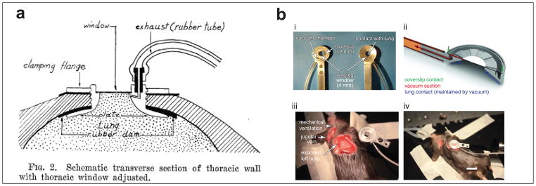
    1. Terry RJ. A THORACIC WINDOW FOR OBSERVATION OF THE LUNG IN A LIVING ANIMAL. Science. 1939;90:43–44. - PubMed
    1. Wagner WW. Pulmonary microcirculatory observations in vivo under physiological conditions. J Appl Physiol. 1969;26:375–377. - PubMed