Review
. 2014:122:103-41.
doi: 10.1016/B978-0-12-420117-0.00003-7.

Gamma-glutamyl transpeptidase: redox regulation and drug resistance

Affiliations
Review

Gamma-glutamyl transpeptidase: redox regulation and drug resistance

Marie H Hanigan. Adv Cancer Res. 2014.

Abstract

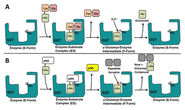
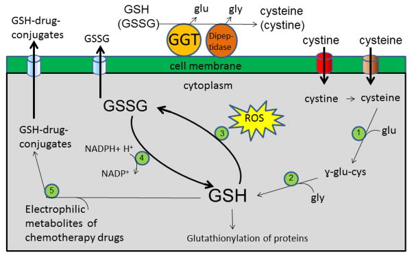
The expression of gamma-glutamyl transpeptidase (GGT) is essential to maintaining cysteine levels in the body. GGT is a cell surface enzyme that hydrolyzes the gamma-glutamyl bond of extracellular reduced and oxidized glutathione, initiating their cleavage into glutamate, cysteine (cystine), and glycine. GGT is normally expressed on the apical surface of ducts and glands, salvaging the amino acids from glutathione in the ductal fluids. GGT in tumors is expressed over the entire cell membrane and provides tumors with access to additional cysteine and cystine from reduced and oxidized glutathione in the blood and interstitial fluid. Cysteine is rate-limiting for glutathione synthesis in cells under oxidative stress. The induction of GGT is observed in tumors with elevated levels of intracellular glutathione. Studies in models of hepatocarcinogenesis show that GGT expression in foci of preneoplastic hepatocytes provides a selective advantage to the cells during tumor promotion with agents that deplete intracellular glutathione. Similarly, expression of GGT in tumors enables cells to maintain elevated levels of intracellular glutathione and to rapidly replenish glutathione during treatment with prooxidant anticancer therapy. In the clinic, the expression of GGT in tumors is correlated with drug resistance. The inhibitors of GGT block GGT-positive tumors from accessing the cysteine in extracellular glutathione. They also inhibit GGT activity in the kidney, which results in the excretion of GSH in the urine and a rapid decrease in blood cysteine levels, leading to depletion of intracellular GSH in both GGT-positive and GGT-negative tumors. GGT inhibitors are being developed for clinical use to sensitize tumors to chemotherapy.

Keywords: Cysteine; Gamma-glutamyl transferase; Gamma-glutamyl transpeptidase; Glutathione.


Figures

Fig. 1
Fig. 1
The van der Waals surface of human GGT with the active site cleft facing the viewer. The large subunit (dark gray) and the small subunit (white) are shown with the catalytic Thr-381 (red) in the deepest part of the small subunit cleft. Four of the seven potential glycosylation sites are seen in this orientation, asparagine 110, 120, 230 and 344, and the basal N-acetyl glucosamine residue, which was identified in the crystal structure at each of these sites, is represented as dark orange van der Waals spheres. An anion-binding site (green) within the small subunit cleft is labeled 1103. This figure was originally published in The Journal of Biological Chemistry. West, M.B., Chen, Y., Wickham, S., Heroux, A., Cahill, K., Hanigan, M.H., Mooers, B.H.M., Novel insights into eukaryotic gamma-glutamyl transpeptidase 1 from the crystal structure of the glutamate bound human enzyme. J. Biol. Chem. 2013; 288(44):31902–31913. © the American Society for Biochemistry and Molecular Biology
Fig. 2
Fig. 2
Gamma-glutamyl bond. The structure of glutathione is shown glutamate (green) bound via a γ-glutamyl bond (arrow) to cysteine (red) and glycine (blue).
Fig. 3
Fig. 3
The Hydrolysis and Transpeptidation Reactions Catalyzed by GGT. (A) The physiological reaction catalyzed by GGT is the hydrolysis of gamma-glutamyl bonds. The hydrolysis reaction is shown with glutathione as the substrate, and shows the release of glutamate which is the product that is measured in the Glutamate Release Assay for GGT activity. (B) The transpeptidation reaction requires high concentration of dipeptide acceptor and is favored at pH 8 and higher. The transpeptidation reaction is shown with L-gamma-glutamyl para-nitroanalide (L-GpNA), the substrate used in the standard biochemical GGT assay, which monitors the release of pNA which is yellow. Adapted from a figure published in Biochemical Journal. Wickham, S., Regan, N., West, M. B., Thai, J., Cook, P. F., Terzyan, S. S., et al. (2013). Inhibition of human gamma-glutamyl transpeptidase: development of more potent, physiologically relevant, uncompetitive inhibitors. Biochem J. 2013; 450(3): 547–557@ The Biochemical Society.
Fig. 4
Fig. 4
Gamma-glutamyl transpeptidase (GGT)-positive tumors cleave extracellular reduced and oxidized glutathione (GSH and GSSG) providing an additional source of cysteine for intracellular GSH synthesis. GGT cleaves glutamate from GSH and GSSG. The cysteinylglycine dipeptides can be cleaved by any of several dipeptidases that are present on the surface of the cell. The glutamate, glycine, cystine and cysteine that are released from GSH and GSSG, are transported into the cell by the standard amino acid transporters. Cystine is taken up by the xc cystine/glutamate antiporter (red). Cysteine is taken up by the ACS transporter (brown). Once inside the cell, cystine is reduced to cysteine by the strongly reducing environment of the cytoplasm. The first step in GSH synthesis is catalyzed by gamma-glutamyl cysteine synthetase (1) and the second step is catalyzed by GSH synthetase (2). Tumors are under redox stress which can be further increased by pro-oxidant therapy. GSH levels are depleted in tumors by several pathways. GSH is oxidized to GSSG as part of an ROS detoxification system that is present in both the cytoplasm and mitochondria. GSH peroxidases (3) catalyze the oxidation of GSH to GSSG. GSSG and be reduced to GSH by GSH reductase (4) and NADPH. However, under extreme oxidative stress GSSG is transported out of the cell by the multidrug resistance protein (MRP) transporters (blue). GSH is also depleted from the cell by binding to the electrophilic metabolites of chemotherapy drugs, which is catalyzed by GSH S-transferases (5). The GSH-conjugates are transported out of the cell by the MRP transporters (blue). Intracellular GSH can also be depleted by binding to intercellular proteins, a process known as protein glutathionylation.

Similar articles

Cited by

References

    1. Ahluwalia GS, Grem JL, Hao Z, Cooney DA. Metabolism and action of amino acid analog anti-cancer agents. Pharmacol Ther. 1990;46(2):243–271. - PubMed
    1. Ahmad S, Okine L, Wood R, Aljian J, Vistica DT. gamma-Glutamyl transpeptidase (gamma-GT) and maintenance of thiol pools in tumor cells resistant to alkylating agents. J Cell Physiol. 1987;131(2):240–246. - PubMed
    1. Anders MW, Dekant W. Glutathione-dependent bioactivation of haloalkenes. Annu Rev Pharmacol Toxicol. 1998;38:501–537. - PubMed
    1. Anderson ME, Bridges RJ, Meister A. Direct evidence for inter-organ transport of glutathione and that the non-filtration renal mechanism for glutathione utilization involves gamma-glutamyl transpeptidase. Biochem Biophys Res Commun. 1980;96(2):848–853. - PubMed
    1. Auman JT, Church R, Lee SY, Watson MA, Fleshman JW, McLeod HL. Celecoxib pre-treatment in human colorectal adenocarcinoma patients is associated with gene expression alterations suggestive of diminished cellular proliferation. Eur J Cancer. 2008;44(12):1754–1760. - PMC - PubMed

Publication types