. 2014 Aug;18(8):1644-54.
doi: 10.1111/jcmm.12351. Epub 2014 Jun 28.

iPSC-derived human mesenchymal stem cells improve myocardial strain of infarcted myocardium

Affiliations

iPSC-derived human mesenchymal stem cells improve myocardial strain of infarcted myocardium

Qingfeng Miao et al. J Cell Mol Med. 2014 Aug.

Abstract

We investigated global and regional effects of myocardial transplantation of human induced pluripotent stem cell (iPSC)-derived mesenchymal stem cells (iMSCs) in infarcted myocardium. Acute myocardial infarction (MI) was induced by ligation of left coronary artery of severe combined immunodeficient mice before 2 × 10(5) iMSCs or cell-free saline were injected into peri-infarcted anterior free wall. Sham-operated animals received no injection. Global and regional myocardial function was assessed serially at 1-week and 8-week by segmental strain analysis by using two dimensional (2D) speckle tracking echocardiography. Early myocardial remodelling was observed at 1-week and persisted to 8-week with global contractility of ejection fraction and fractional area change in saline- (32.96 ± 14.23%; 21.50 ± 10.07%) and iMSC-injected (32.95 ± 10.31%; 21.00 ± 7.11%) groups significantly depressed as compared to sham control (51.17 ± 11.69%, P < 0.05; 34.86 ± 9.82%, P < 0.05). However, myocardial dilatation was observed in saline-injected animals (4.40 ± 0.62 mm, P < 0.05), but not iMSCs (4.29 ± 0.57 mm), when compared to sham control (3.74 ± 0.32 mm). Furthermore, strain analysis showed significant improved basal anterior wall strain (28.86 ± 8.16%, P < 0.05) in the iMSC group, but not saline-injected (15.81 ± 13.92%), when compared to sham control (22.18 ± 4.13%). This was corroborated by multi-segments deterioration of radial strain only in saline-injected (21.50 ± 5.31%, P < 0.05), but not iMSC (25.67 ± 12.53%), when compared to sham control (34.88 ± 5.77%). Improvements of the myocardial strain coincided with the presence of interconnecting telocytes in interstitial space of the infarcted anterior segment of the heart. Our results show that localized injection of iMSCs alleviates ventricular remodelling, sustains global and regional myocardial strain by paracrine-driven effect on neoangiogenesis and myocardial deformation/compliance via parenchymal and interstitial cell interactions in the infarcted myocardium.

Keywords: cell therapy; myocardial compliance; myocardial strain; telocytes; tissue deformation.


Figures

Fig. 1
Fig. 1
Short-axis and long-axis views of speckle tracking echocardiography. (A) Six-segment view from short axis of a mouse heart. (B) Six-segment view from long axis of a mouse heart. Ant.: anterior; post.: posterior; Inf.: inferior.
Fig. 2
Fig. 2
Global peak strain analysis of LV systolic deformations at baseline and follow-up. SAX: short-axis view; LAX: long-axis view.
Fig. 3
Fig. 3
Myocardial localization of transplanted human iMSC in the anterior segment of left ventricle. (A) Six-segment view of transverse sectioned mouse heart injected with iMSCs. (B) Human-specific nuclear staining of Ku80 in iMSCs in peri-infarcted zone of anterior segment bordering lateral wall. (C) Human-specific nuclear staining of Ku80 in iMSCs in peri-infarcted zone of anterior segment next to infarcted wall. (D) Identification of human Ku80 stained iMSCs in the infarcted zone of anterior wall. (EG) Magnified views of boxed region in B, C and D. (H) Transverse sectioned view of saline-injected mouse heart. (J) Magnified view of infarct border in H. (I) Transverse sectioned view of sham-operated mouse heart. (K) Magnified view of injured border in I. Scale bar: 1 mm (A, H and I); 20 μm (for all others).
Fig. 4
Fig. 4
Interstitial localization of transplanted human iMSCs in myocardium. (A) Presence of human-specific nuclear staining of Ku80 iMSCs in the interstitial space of cardiac muscle. (BD) Magnified view of boxed region in A, showing Ku80 stained iMSCs located mainly in the interstitial space (demarcated line) of α-actinin (green) stained cardiac muscle. (E) Presence of human-specific nuclear staining of Ku80 iMSCs in fibrous/collagenous region of myocardium. (FH) Magnified view of boxed region in E, showing Ku80 stained human iMSCs (arrows) located in peri-vascular space of α-smooth muscle actin (SMA) stained microvessels; scale bar: 50 μm.
Fig. 5
Fig. 5
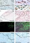
Presence of CD34 stained interstitial cells in the infarct zone. (A) Interstitial cells with long cellular processes that stained positive for CD34 that resided longitudinally in the collagen-rich and remodelling infarct zone of iMSC transplanted heart. (B and C) Magnified view of CD34 stained cells with thin and long cellular processes resembled podomers (arrows) and podom (arrowhead) of myocardial telocytes in the interstitial space. (D) Presence of human-specific nuclear staining of Ku80 iMSCs and interstitial cells in peri-infarct zone. (EG) Magnified view of boxed region in D, showing Ku80 and CD34 double-stained human telocyte (arrow) in close proximity with CD34 stained (but human Ku80 negative) resident telocyte (*). (H) Absence of CD34 stained interstitial cells in the infarct zone of saline-injected mouse heart. (I) Absence of CD34 stained interstitial cells in the injured zone of sham-operated mouse heart. Dotted line demarcates intact myocardium from infarct zone. Scale bar: 50 μm (A, DI); 10 μm (B and C).
Fig. 6
Fig. 6
Infarct staining with Masson's trichrome staining. (A). Sham-operated mouse heart showing epicardial injury with limited fibrotic response and transmural anterior infarct with wall thinning and myocardial fibrosis in saline-injected and iMSC-injected mouse heart. (B) Quantitative estimation of infarct size at 8-week after infarction; scale bar: 1 mm.
Fig. 7
Fig. 7
Microvascular neoangiogenesis at 8-week after myocardial infarction. (A) Immunofluorescent staining for von Willebrand (vWF) and a-smooth muscle actin (SMA) in left ventricle post-infarction. (B) Vascular density counts of vWF stained microvasculature in the left ventricle. (C) Vascular maturity estimated from vWF/SMA co-stained microvessels in the left ventricle; scale bar: 50 μm.

Similar articles

Cited by

References

    1. Miettinen JA, Ylitalo K, Hedberg P, et al. Determinants of functional recovery after myocardial infarction of patients treated with bone marrow-derived stem cells after thrombolytic therapy. Heart. 2010;96:362–7. - PubMed
    1. Hare JM, Traverse JH, Henry TD, et al. A randomized, double-blind, placebo-controlled, dose-escalation study of intravenous adult human mesenchymal stem cells (prochymal) after acute myocardial infarction. J Am Coll Cardiol. 2009;54:2277–86. - PMC - PubMed
    1. Chen SL, Fang WW, Qian J, et al. Improvement of cardiac function after transplantation of autologous bone marrow mesenchymal stem cells in patients with acute myocardial infarction. Chin Med J (Engl) 2004;117:1443–8. - PubMed
    1. Mehta A, Shim W. Cardiac stem cell therapy: stemness or commitment? Cell Transplant. 2013;22:1–14. - PubMed
    1. Zheng Y, Cretoiu D, Yan G, et al. Comparative proteomic analysis of human lung telocytes with fibroblasts. J Cell Mol Med. 2014;18:568–89. - PMC - PubMed

Publication types