Review
. 2014 Jul;10(3):407-18.
doi: 10.1016/j.hfc.2014.04.008. Epub 2014 May 22.

Phenotypic spectrum of heart failure with preserved ejection fraction

Affiliations
Review

Phenotypic spectrum of heart failure with preserved ejection fraction

Sanjiv J Shah et al. Heart Fail Clin. 2014 Jul.

Abstract

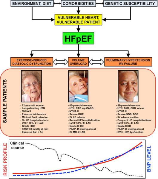
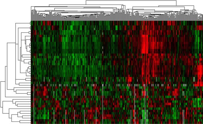
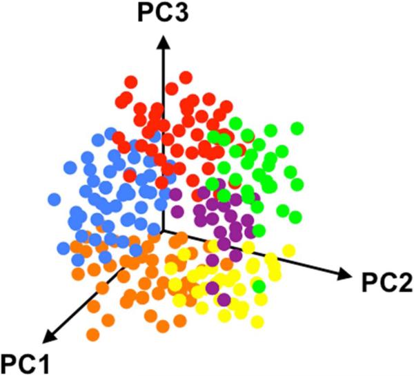
Heart failure with preserved ejection fraction (HFpEF) is a heterogeneous syndrome, with several underlying etiologic and pathophysiologic factors. The heterogeneity of the HFpEF syndrome may explain why (1) diagnosing and treating HFpEF is so challenging and (2) clinical trials in HFpEF have failed thus far. Here we describe 4 ways of categorizing HFpEF based on pathophysiology, clinical/etiologic subtype, type of clinical presentation, and quantitative phenomics (phenomapping analysis). Regardless of the classification method used, improved phenotypic characterization of HFpEF, and matching targeted therapies with specific HFpEF subtypes, will be a critical step towards improving outcomes in this increasingly prevalent syndrome.

Keywords: Classification; Clinical trials; Etiology; Heart failure with preserved ejection fraction; Pathophysiology; Phenomics; Phenotype.


Figures

Figure 1
Figure 1. Color Doppler Echocardiography from a Patient with Heart Failure and Preserved Ejection Fraction Showing Multiple Moderate Valvular Lesions
Caption: Left panel: apical 4-chamber view of the right heart showing moderate tricuspid regurgitation; middle panel: apical 4-chamber view of the left heart showing moderate mitral regurgitation; right panel: apical 3-chamber view showing moderate aortic regurgitation. From Oktay AA, Shah SJ. Heart failure with preserved ejection fraction. In Levine G, ed. Color Atlas of Clinical Cardiology. New Delhi: Jaypee Medical Publishers, 2014; with permission.
Figure 2
Figure 2. Relationship Between Heart Failure Risk Factor Combinations and Heart Failure Phenotypic Heterogeneity
Caption: Multiple different risk factors can lead to different patterns and types of the HF syndrome. Particular combinations of risk factors (i.e., a roll of dice) may lead to different types of HF phenotypes (i.e., a particular hand of cards). HF—heart failure; CO—cardiac output; PCWP—pulmonary capillary wedge pressure
Figure 3
Figure 3. Illustration of a Targeted Diagnostic Approach (e.g., Cancer) versus a One-Size-Fits-All Approach (e.g., Heart Failure)
Caption: Treatment of cancer has benefitted from an increasingly targeted approach whereas current heart failure treatment is more generalized with few exceptions (e.g., cardiac resynchronization therapy). HF—heart failure; ECG—electrocardiography; PEX—physical examination; LVEF—left ventricular ejection fraction
Figure 4
Figure 4. Multiple Pathophysiologic Contributors to the Heart Failure with Preserved Ejection Fraction Syndrome
Caption: HFpEF—heart failure with preserved ejection fraction; RV—right ventricle. From Oktay AA, Shah SJ. Heart failure with preserved ejection fraction. In Levine G, ed. Color Atlas of Clinical Cardiology. New Delhi: Jaypee Medical Publishers, 2014; with permission.
Figure 5
Figure 5. Theoretical Schema of Heart Failure With Preserved Ejection Fraction Patient Types With Sample Patients, Risk Profiles, and Matched Therapies
Caption: AR—aortic regurgitation; ARNI—angiotensin receptor/neprilysin inhibitor; BNP—B-type natriuretic peptide; CABG—coronary artery bypass grafting; CAD—coronary artery disease; CKD—chronic kidney disease; DD—diastolic dysfunction; DM2—type 2 diabetes mellitus; DOE—dyspnea on exertion; E/e’—ratio of early mitral inflow to early mitral annular diastolic tissue velocity; HF—heart failure; HTN—hypertension; If—inward “funny” channel; LAE—left atrial enlargement; LVEF—left ventricular ejection fraction; MR—mitral regurgitation; MRA—mineralocorticoid receptor antagonist; NYHA—New York Heart Association; PASP— pulmonary artery systolic pressure; PDE5—phosphodiesterase-5; RV—right ventricular; RVH— right ventricular hypertrophy; SOB—shortness of breath; s/p—status post. From Shah SJ. Matchmaking for the optimization of clinical trials of heart failure with preserved ejection fraction: no laughing matter. J Am Coll Cardiol 2013;62(15):1339-1342; with permission.
Figure 6
Figure 6. Comprehensive Echocardiographic Phenotypic Analysis of Heart Failure with Preserved Ejection Fraction
Caption: Comprehensive echocardiography, including two dimensional, Doppler, tissue Doppler, and speckle tracking, allows for detailed phenotypic analysis of cardiac structure, function, and mechanics in patients with heart failure with preserved ejection fraction. The figure shows examples of information that can be obtained from the apical 4-chamber view. Clockwise from the top: speckle-tracking echocardiography for assessment of LV regional and global longitudinal strain (early diastolic strain rate can also be obtained in this view). Mitral inflow and tissue Doppler imaging of the septal and lateral mitral annulus provide information on LV diastolic function grade and estimated LV filling pressure (E/e’ ratio), along with assessment of longitudinal systolic (s’) and atrial (a’) function. Speckle-tracking analysis of LA function provides peak LA contractile function (peak negative longitudinal LA strain) and LA reservoir function (peak positive longitudinal LA strain). Tricuspid annular plane systolic function (TAPSE) and basal RV free wall peak longitudinal tissue Doppler velocity (RV s’) provide information on longitudinal RV function, as does speckle tracking echocardiography of the RV (not shown). Finally, analysis of the tricuspid regurgitant jet Doppler profile, when added to the estimated RA pressure, provides an estimate of the PA systolic pressure. Additional data available from the apical 4-chamber view include assessment of LV volumes and ejection fraction, LA volume, and RV size and global systolic function (e.g., RV fractional area change). LV—left ventricular; LA—left atrial; PA—pulmonary artery; RV—right ventricular; RA—right atrial; A4C—apical 4-chamber From Butler J, Fonarow G, Zile MR, et al. Developing Therapies for Heart Failure with Preserved Ejection Fraction: Current State and Future Directions. JACC Heart Fail 2014 (in press); with permission.
Figure 7
Figure 7. Sample Phenotypic Heat Map (“Pheno-Map”) Developed from Hierarchical Cluster Analysis of Quantitative Echocardiographic Data
Caption: The rows in the heat map correspond to the various quantitative echocardiographic phenotypes (e.g., septal wall thickness, ejection fraction, early diastolic [e’] tissue velocity, etc.), while the columns represent individual patients. Red = increased values; green = decreased values. The dendrogram across the top of the heat map is a tree diagram that demonstrates the clustering of patients; the dendrogram on the left side of the heat map illustrates clustering of phenotypes.
Figure 8
Figure 8. Three-Dimensional Principal Components Analysis Plot
Caption: In this theoretical example, patients are grouped based on 3 principal components (PC1, PC2, and PC3) in 3 dimensions. Each color represents a group of patients that correspond to a particular cluster based on phenotypic similarities.

References

    1. Yancy CW, Jessup M, Bozkurt B, et al. 2013 ACCF/AHA Guideline for the Management of Heart Failure: A Report of the American College of Cardiology Foundation/American Heart Association Task Force on Practice Guidelines. J Am Coll Cardiol. 2013 - PubMed
    1. Oktay AA, Shah SJ. Diagnosis and Management of Heart Failure with Preserved Ejection Fraction: 10 Key Lessons. Curr Cardiol Rev. 2013 - PMC - PubMed
    1. Oktay AA, Rich JD, Shah SJ. The emerging epidemic of heart failure with preserved ejection fraction. Curr Heart Fail Rep. 2013;10(4):401–410. - PMC - PubMed
    1. Go AS, Mozaffarian D, Roger VL, et al. Heart disease and stroke statistics--2013 update: a report from the American Heart Association. Circulation. 2013;127(1):e6–e245. - PMC - PubMed
    1. Shah AM, Pfeffer MA. The many faces of heart failure with preserved ejection fraction. Nat Rev Cardiol. 2012;9(10):555–556. - PubMed

Publication types

MeSH terms

Substances