

Traumatic pseudoaneurysm of the middle meningeal artery with an arteriovenous fistula on a non-fractured site
- PMID: 24976099
- PMCID: PMC4178773
- DOI: 10.15274/INR-2014-10025
Traumatic pseudoaneurysm of the middle meningeal artery with an arteriovenous fistula on a non-fractured site
Abstract
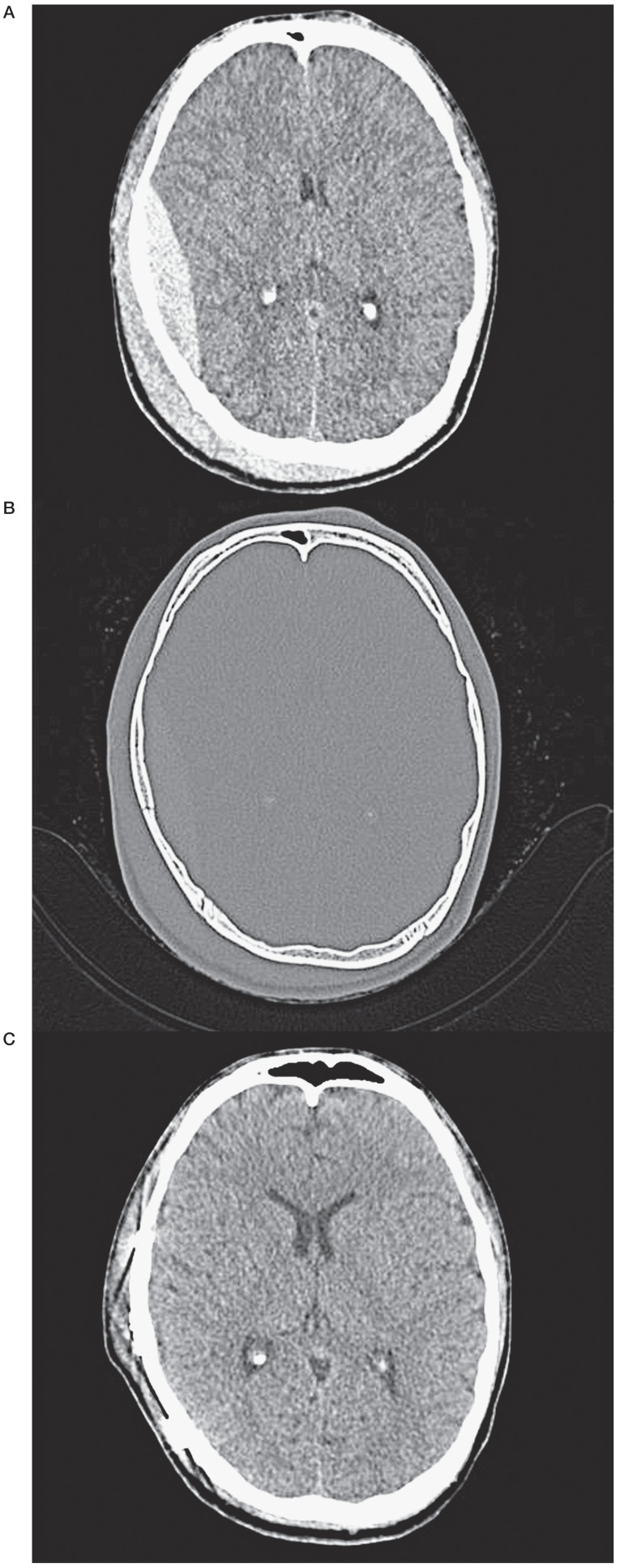
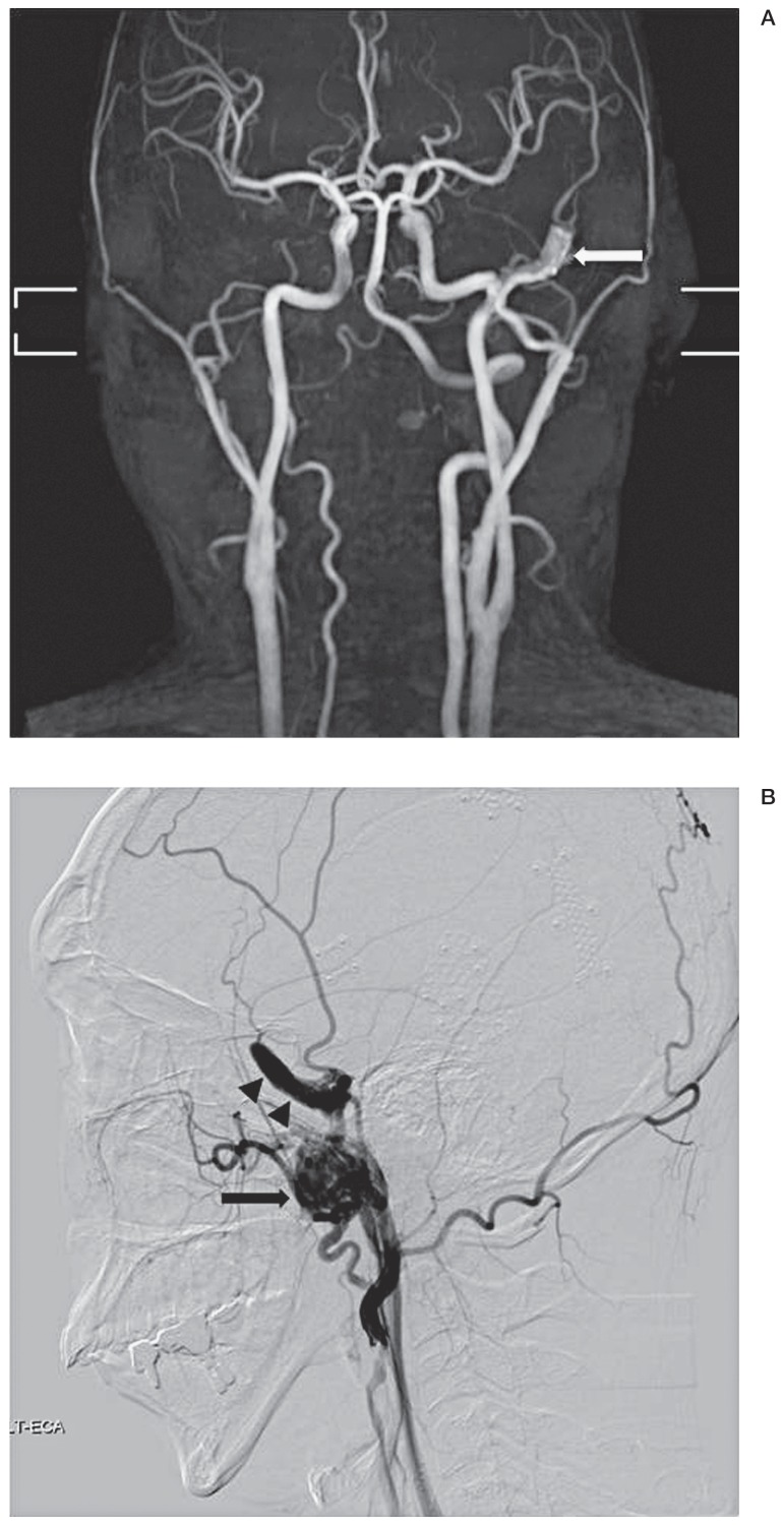
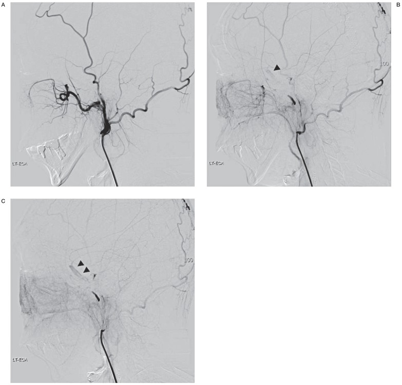
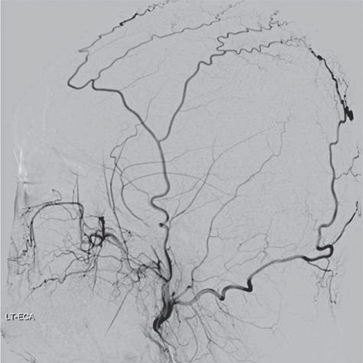
We describe a rare case of a combined traumatic pseudoaneurysm and arteriovenous fistula (AVF) of the middle meningeal artery (MMA) on a non-fractured site. A 24-year-old man was admitted to our hospital with head trauma. He underwent a craniotomy and removal of an epidural hematoma on the right side. Twenty-five days later, he complained of pulsatile tinnitus on the left non-fractured side. Angiography revealed a markedly dilated proximal MMA with flow shunting to the pterygoid plexus. We performed proximal occlusion on the proximal MMA for the traumatic pseudoaneurysm and the AVF of the MMA using coils. Although immediate angiography showed retrograde contrast filling from the collateral vessels into the distal part of the pseudoaneurysm, follow-up angiography revealed that the lesion had successfully disappeared.
Keywords: arteriovenous fistula; head trauma; middle meningeal artery; pseudoaneurysm.
Figures
References
-
- Jussen D, Wiener E, Vajkoczy P, et al. Traumatic middle meningeal artery pseudoaneurysms: diagnosis and endovascular treatment of two cases and review of the literature. Neuroradiology. 2012;54(10):1133–1136. doi: 10.1007/s00234-011-1003-7. - DOI - PubMed
-
- Rennert J, Seiz M, Nimsky C, et al. Endovascular treatment of traumatic high flow dural arterio-venous fistula involving the middle meningeal artery and facial veins. Rontgenpraxis. 2008;56(5):164–168. doi: 10.1016/j.rontge.2006.10.002. - DOI - PubMed
-
- Lim DH, Kim TS, Joo SP, et al. Intracerebral hematoma caused by ruptured traumatic pseudoaneurysm of the middle meningeal artery: a case report. J Korean Neurosurg Soc. 2007;42(5):416–418. doi: 10.3340/jkns.2007.42.5.416. - DOI - PMC - PubMed
-
- Freckmann N, Sartor K, Herrmann HD. Traumatic arteriovenous fistulae of the middle meningeal artery and neighbouring veins or dural sinuses. Acta Neurochir (Wien) 1981;55(3-4):273–281. doi: 10.1007/BF01808443. - DOI - PubMed
-
- Kawaguchi T, Kawano T, Kaneko Y, et al. Traumatic lesions of the bilateral middle meningeal arteries--case report. Neurol Med Chir (Tokyo) 2002;42(5):221–223. doi: 10.2176/nmc.42.221. - DOI - PubMed
Publication types
MeSH terms
LinkOut - more resources
Full Text Sources
Other Literature Sources
