

New vascular classification of port-wine stains: improving prediction of Sturge-Weber risk
- PMID: 24976116
- PMCID: PMC4284033
- DOI: 10.1111/bjd.13203
New vascular classification of port-wine stains: improving prediction of Sturge-Weber risk
Abstract
Background: Facial port-wine stains (PWSs) are usually isolated findings; however, when associated with cerebral and ocular vascular malformations they form part of the classical triad of Sturge-Weber syndrome (SWS).
Objectives: To evaluate the associations between the phenotype of facial PWS and the diagnosis of SWS in a cohort with a high rate of SWS.
Methods: Records were reviewed of all 192 children with a facial PWS seen in 2011-13. Adverse outcome measures were clinical (seizures, abnormal neurodevelopment, glaucoma) and radiological [abnormal magnetic resonance imaging (MRI)], modelled by multivariate logistic regression.
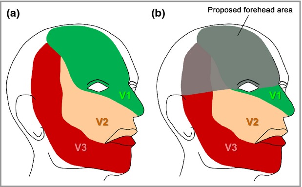
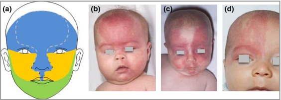
Results: The best predictor of adverse outcomes was a PWS involving any part of the forehead, delineated at its inferior border by a line joining the outer canthus of the eye to the top of the ear, and including the upper eyelid. This involves all three divisions of the trigeminal nerve, but corresponds well to the embryonic vascular development of the face. Bilateral distribution was not an independently significant phenotypic feature. Abnormal MRI was a better predictor of all clinical adverse outcome measures than PWS distribution; however, for practical reasons guidelines based on clinical phenotype are proposed.
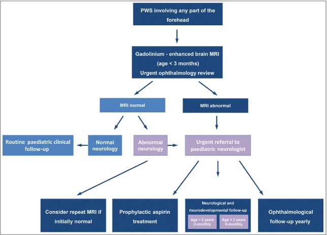
Conclusions: Facial PWS distribution appears to follow the embryonic vasculature of the face, rather than the trigeminal nerve. We propose that children with a PWS on any part of the 'forehead' should have an urgent ophthalmology review and a brain MRI. A prospective study has been established to test the validity of these guidelines.
© The Authors. British Journal of Dermatology published by John Wiley & Sons Ltd on behalf of British Association of Dermatologists.
Figures
Comment in
-
Patterns of vascular birthmarks: questions and clues.Br J Dermatol. 2014 Oct;171(4):693-4. doi: 10.1111/bjd.13316. Br J Dermatol. 2014. PMID: 25319426 No abstract available.
-
Nonrandom distribution of facial tufted angiomas.Int J Dermatol. 2022 May;61(5):e193-e194. doi: 10.1111/ijd.15808. Epub 2021 Jul 15. Int J Dermatol. 2022. PMID: 34263941 No abstract available.
References
-
- Sturge WA. A case of partial epilepsy apparently due to a lesion of one of the vasomotor centres of the brain. Trans Clin Soc Lond. 1879;12:162–7.
-
- Kalischer S. Ein Fall von Telangiektasie (Angiom) des Gesichts und der weichen Hirnhäute. Arch Psychiatr Nervenkrankheiten Berl. 1901;34:171–80. (in German)
-
- Piram M, Lorette G, Sirinelli D, et al. Sturge-Weber syndrome in patients with facial port-wine stain. Pediatr Dermatol. 2012;29:32–7. - PubMed
Publication types
MeSH terms
LinkOut - more resources
Full Text Sources
Other Literature Sources
