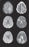
The etiology of ring lesions on diffusion-weighted imaging
- PMID: 24976194
- PMCID: PMC4202903
- DOI: 10.15274/NRJ-2014-10036
The etiology of ring lesions on diffusion-weighted imaging
Abstract
This study describes a series of cases and reviews the literature on cases of ring lesion on diffusion-weighted imaging to better appreciate the spectrum of disease associated with this neuroimaging finding. We retrospectively reviewed the MR studies of 15 patients with ring pattern lesions on diffusion-weighted imaging from an inpatient Neurology service of a tertiary care center seen over a ten-year period, and reviewed cases in the literature. Thirty-one cases, including 15 new patients, comprise the study group. Immunocompromised patients accounted for 38% of patients with ring lesions on diffusion-weighted imaging with cerebral aspergillosis in five patients, progressive multifocal leukoencephalopathy in three, primary CNS lymphoma in two, cerebral toxoplasmosis in one, and resolving cerebral hematoma in one. In the immunocompetent group demyelinating lesions including multiple sclerosis, acute disseminated encephalomyelitis, Balo's concentric sclerosis and acute necrotizing encephalitis, were seen in 11 patients, vascular etiology in four and neoplastic in three patients, two primary and one metastatic and pyogenic brain abscess in one. Ring lesions on diffusion-weighted imaging are associated with a spectrum of disease not previously considered. Immunocompromised patients accounted for almost one-half while demyelinating conditions in the immunocompetent patients were most common overall.
Keywords: MRI; demyelinating disease; diffusion-weighted imaging; immunocompromised; ring lesion.
Figures


References
-
- Schaefer PW, Grant E, Gonzalez G. Diffusion-weighted MR imaging of the brain. Radiology. 2000;217(2):331–345. doi: 10.1148/radiology.217.2.r00nv24331. - DOI - PubMed
-
- Finelli PF, Gleeson E, Ciesielski T, et al. Diagnostic role of target lesion on diffusion-weighted imaging. A case of cerebral aspergillosis and review of the literature. Neurologist. 2010;16(6):364–367. doi: 10.1097/NRL.0b013e3181b4700. - DOI - PubMed
-
- Finelli PF. Primary CNS lymphoma in myasthenic on long-term azathioprine. J Neurooncol. 2005;74(1):91–92. doi: 10.1007/s11060-004-5676-1. - DOI - PubMed
-
- Finelli PF, Uphoff DF. Ring Lesion on DWI. Arch Neurol. 2011;68(11):1474–1475. doi: 10.1001/archneurol.2011.612. - DOI - PubMed
-
- Finelli PF, Naik K, DiGiuseppe JA, et al. Primary lymphoma of CNS, mycophenolate mofetil and lupus. Lupus. 2006;15(12):886–888. doi: 10.1177/0961203306071431. - DOI - PubMed
Publication types
MeSH terms
LinkOut - more resources
Full Text Sources
Other Literature Sources
Medical

