

Lentiform fork sign: a magnetic resonance finding in a case of acute metabolic acidosis
- PMID: 24976195
- PMCID: PMC4202895
- DOI: 10.15274/NRJ-2014-10041
Lentiform fork sign: a magnetic resonance finding in a case of acute metabolic acidosis
Abstract
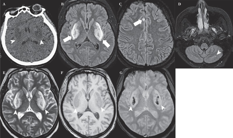
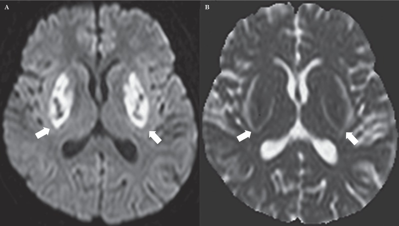
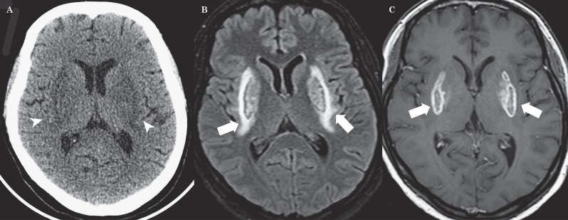
We report a 33 year-old woman addicted to chronic unspecified solvents abuse with stupor, respiratory disorders, tetraplegia and severe metabolic acidosis. On admission an unenhanced cranial CT scan showed symmetrical hypodensities of both lentiform nuclei. MR imaging performed 12 hours after stupor demonstrates bilateral putaminal hemorrhagic necrosis, bilateral external capsule, corona radiata and deep cerebellar hyperintensities with right cingulate cortex involvement. DWI reflected bilateral putaminal hyperintensities with restricted water diffusion as to citotoxic edema and development of vasogenic edema in the external capsule recalling a fork. On day twenty, after specific treatments MRI demonstrated a bilateral putaminal marginal enhancement. Bilateral putaminal necrosis is a characteristic but non-specific radiological finding of methanol poisoning. Lentiform Fork sign is a rare MRI finding reported in literature in 22 patients with various conditions characterized by metabolic acidosis. Vasogenic edema may be due to the differences in metabolic vulnerability between neurons and astrocytes. We postulate that metabolic acidosis could have an important role to generate this sign.
Keywords: MRI; diffusion-weighted imaging; lentiform fork sign; metabolic acidosis; putaminal necrosis.
Figures
References
-
- Albin RL. Basal ganglia neurotoxins. Neurol Clin. 2000;18(3):665–680. doi: 10.1016/S0733-8619(05)70217-6. - DOI - PubMed
-
- Beltz EE, Mullins ME. Radiological reasoning: hyperintensity of the basal ganglia and cortex on FLAIR and diffusion-weighted imaging. Am J Roentgenol. 2010;195(3 Suppl):S1–8. (Quiz S9-11). doi: 10.2214/AJR.07.7089. - DOI - PubMed
-
- Kumar G, Goyal MK. Lentiform fork sign: a unique MRI picture. Is metabolic acidosis responsible? Clin Neurol Neurosurg. 2010;112(9):805–812. doi: 10.1016/j.clineuro.2010.06.006. - DOI - PubMed
-
- Wang HC, Cheng SJ. The syndrome of acute bilateral basal ganglia lesions in diabetic uremic patients. J Neurol. 2003;250(8):948–955. doi: 10.1007/s00415-003-1122-0. - DOI - PubMed
-
- Yoon CH, Seok JI, Lee DK, et al. Bilateral basal ganglia and unilateral cortical involvement in a diabetic uremic patient. Clin Neurol Neurosurg. 2009;111(5):477–479. doi: 10.1016/j.clineuro.2009.01.007. - DOI - PubMed
Publication types
MeSH terms
LinkOut - more resources
Full Text Sources
Other Literature Sources
Medical
