

Case Reports
doi: 10.15274/NRJ-2014-10047.
Epub 2014 Jun 17.
Giant arachnoid granulations mimicking pathology. A report of three cases
Affiliations
- PMID: 24976198
- PMCID: PMC4202885
- DOI: 10.15274/NRJ-2014-10047
Item in Clipboard
Case Reports
Giant arachnoid granulations mimicking pathology. A report of three cases
Neuroradiol J.
2014 Jun.
Abstract
We describe three cases of incidentally found lesions in the dural venous sinuses on magnetic resonance imaging, that other had erroneously considered pathological entities. We made the diagnosis of giant arachnoid granulations. The differential diagnosis with thrombosis or intrasinusal tumoral lesions was easily made on the basis of three typical radiological features of the granulations: the hyperintensity of the lesions on FLAIR, a blood vessel within the lesion and bone erosion.
Keywords: CT; MR; giant arachnoid granulations.
Figures
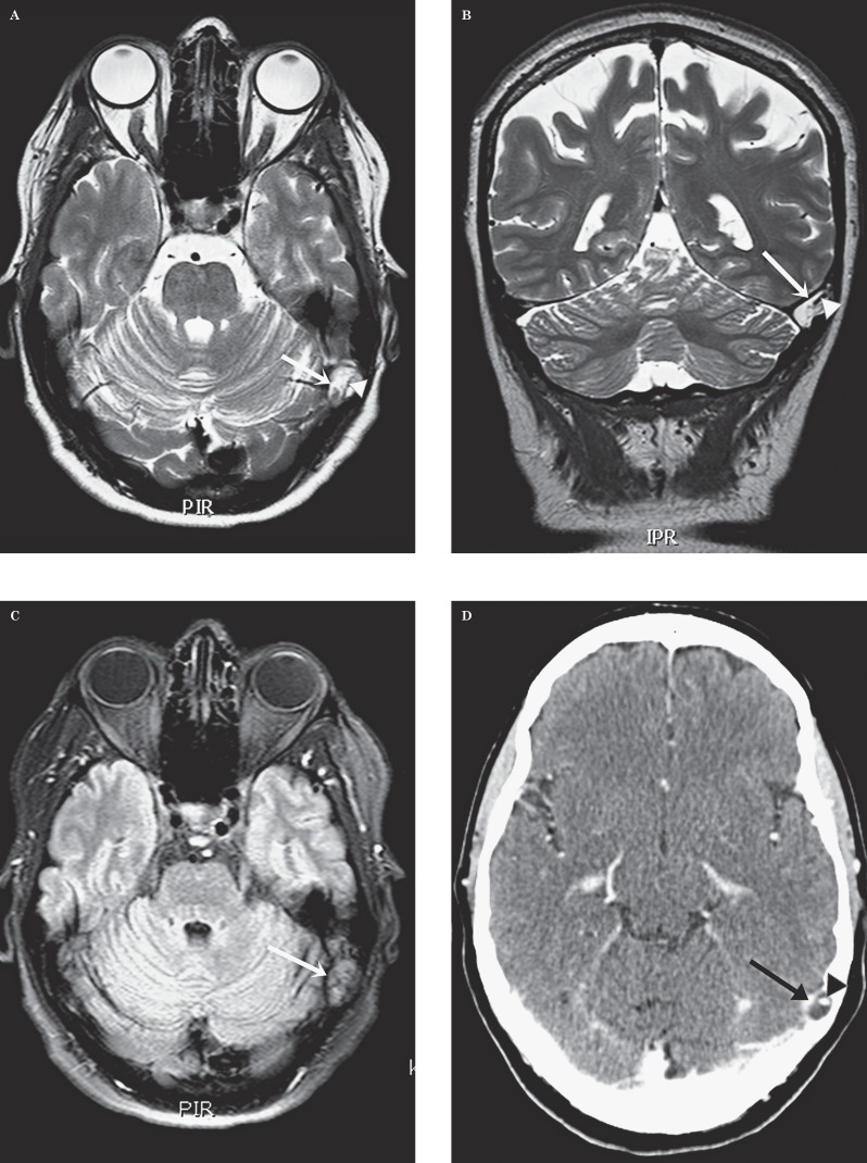
Patient 1. Giant arachnoid granulation in the left transverse sinus. Transverse (A) and coronal (B) T2-weighted images. An ovalar hyperintense structure (arrow) can be seen in the left transverse sinus at the transition to the sigmoid sinus. Intralesional flow void (arrowhead). C) Transverse FLAIR. There is attenuation of the content of this “lesion” (arrow) that remains hyperintense. Transverse (D) and coronal (E) enhanced CT. The “lesion” (arrow) is hypodense with a central enhancing vascular structure (arrowhead). E) Coronal CT with bone windows. Note the scalloping of the overlying bone (arrow).
Patient 1. Giant arachnoid granulation in the left transverse sinus. Transverse (A) and coronal (B) T2-weighted images. An ovalar hyperintense structure (arrow) can be seen in the left transverse sinus at the transition to the sigmoid sinus. Intralesional flow void (arrowhead). C) Transverse FLAIR. There is attenuation of the content of this “lesion” (arrow) that remains hyperintense. Transverse (D) and coronal (E) enhanced CT. The “lesion” (arrow) is hypodense with a central enhancing vascular structure (arrowhead). E) Coronal CT with bone windows. Note the scalloping of the overlying bone (arrow).
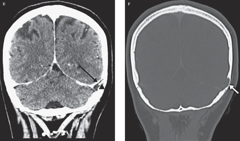
Patient 2. Giant arachnoid granulation in the right transverse sinus. A) Transverse FLAIR. Note the hyperintense “lesion” (arrow) in the left transverse sinus at the transition to the sigmoid sinus. Central flow void pointing to a vascular structure (arrowhead). B) Transverse gadolinium-enhanced image. The hypointense ovalar lesion (arrow) in the transverse sinus is confirmed. The vascular structure less well visible due to volume-averaging.
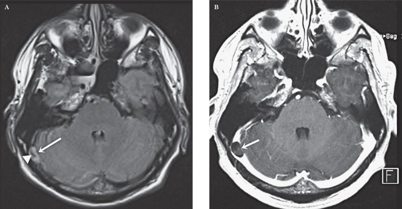
Patient 3. Giant arachnoid granulation in the superior sagittal sinus. Transverse (A) and sagittal (B) T2-weighted image. Huge ovalar hyperintense structure (arrow) within the superior sagittal sinus in the under third. Intralesional vascular flow-void (arrowhead). C) Sagittal gadolinium-enhanced image. Confirmation of the granulation (arrow) and the vascular structure (arrowhead).
References
-
- Leach JL, Meyer K, Jones BV, et al. A large arachnoid granulations involving the dorsal superior sagittal sinus: findings on MR imaging and MR venography. Am J Neuroradiol. 2008;29:1335–1339. doi: 10.3174/ajnr.A1093. - DOI - PMC - PubMed
-
- Trimble CR, Harnsberger HR, Castillo M, et al. "Giant" arachnoid granulations just like CSF? Not. Am J Neuroradiol. 2010;31:1724–1728. doi: 10.3174/ajnr.A2157. - DOI - PMC - PubMed
Publication types
MeSH terms
LinkOut - more resources
Full Text Sources
Other Literature Sources
Medical
