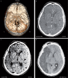
Letter to the Editor. CT angiography source-images and CT perfusion: are they complementary tools for ischemic stroke evaluation? Response to: reliability of CT perfusion in the evaluation of ischaemic penumbra
- PMID: 24976206
- PMCID: PMC4202902
- DOI: 10.15274/NRJ-2014-10043
Letter to the Editor. CT angiography source-images and CT perfusion: are they complementary tools for ischemic stroke evaluation? Response to: reliability of CT perfusion in the evaluation of ischaemic penumbra
Keywords: CT angiography; CT perfusion; source-images ischemic stroke.
Figures

Comment in
-
Response to letter to the editor. "CT angiography source-images and CT perfusion: are they complementary tools for ischemic stroke evaluation?".Neuroradiol J. 2014 Jun;27(3):368. doi: 10.15274/NRJ-2014-10044. Epub 2014 Jun 17. Neuroradiol J. 2014. PMID: 24976207 Free PMC article. No abstract available.
Comment on
-
Reliability of CT perfusion in the evaluation of the ischaemic penumbra.Neuroradiol J. 2014 Feb;27(1):91-5. doi: 10.15274/NRJ-2014-10010. Epub 2014 Feb 24. Neuroradiol J. 2014. PMID: 24571838 Free PMC article.
References
-
- Alves JE, Carneiro A, Xavier J. Reliability of CT perfusion in the evaluation of the ischaemic penumbra. Neuroradiol J. 2014;27(1):91–95. doi: 10.15274/NRJ-2014-10010. - DOI - PMC - PubMed
-
- Schramm P, Schellinger PD, Fiebach JB, et al. Comparison of CT and CT angiography source images with diffusion-weighted imaging in patients with acute stroke within 6 hours after onset. Stroke. 2002;33(10):2426–2432. doi: 10.1161/01.STR.0000032244.03134.37. - DOI - PubMed
-
- Lin K, Rapalino O, Law M, et al. Accuracy of the Alberta Stroke Program Early CT Score during the first 3 hours of middle cerebral artery stroke: comparison of noncontrast CT, CT angiography source images, and CT perfusion. Am J Neuroradiol. 2008;9(5):931–936. doi: 10.3174/ajnr.A0975. - DOI - PMC - PubMed
-
- Kamalian S, Kamalian S, Maas MB, et al. CT cerebral blood flow maps optimally correlate with admission diffusion-weighted imaging in acute stroke but thresholds vary by postprocessing platform. Stroke. 2011;42(7):1923–1928. doi: 10.1161/STROKEAHA.110.610618. - DOI - PMC - PubMed
Publication types
MeSH terms
LinkOut - more resources
Full Text Sources
Other Literature Sources
Medical

