. 2014 Jul 16;83(2):444-454.
doi: 10.1016/j.neuron.2014.05.028. Epub 2014 Jun 26.

A role for Tac2, NkB, and Nk3 receptor in normal and dysregulated fear memory consolidation

Affiliations

A role for Tac2, NkB, and Nk3 receptor in normal and dysregulated fear memory consolidation

Raül Andero et al. Neuron. .

Abstract

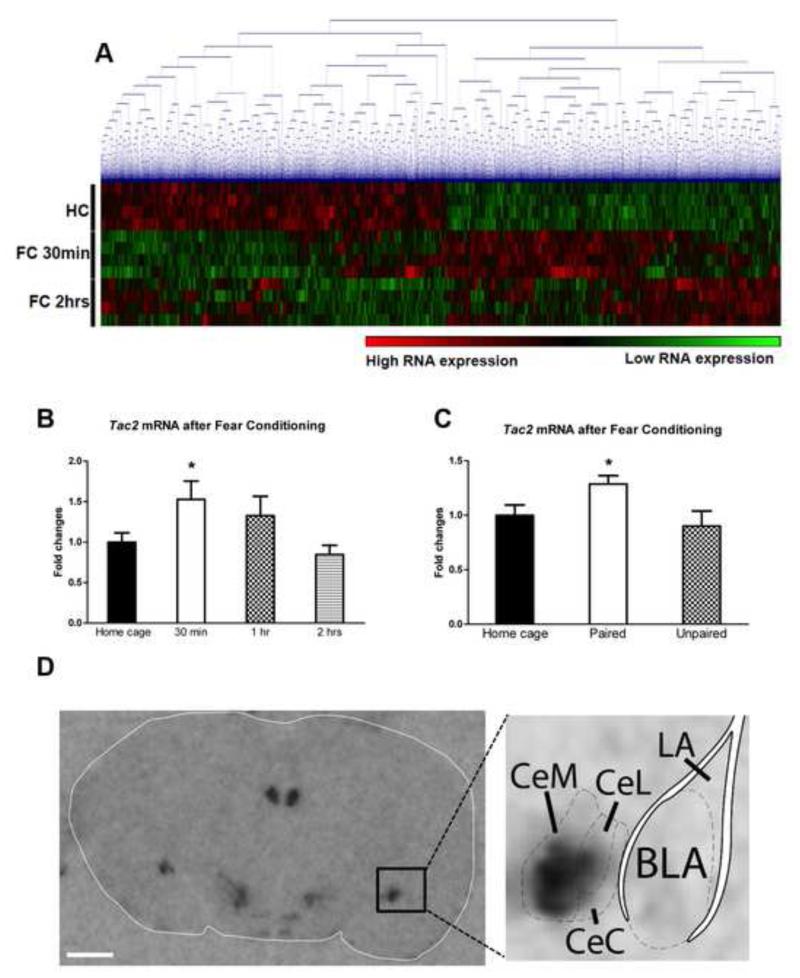
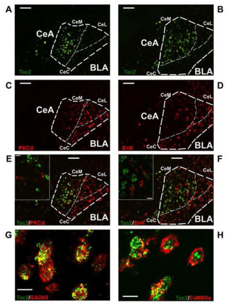
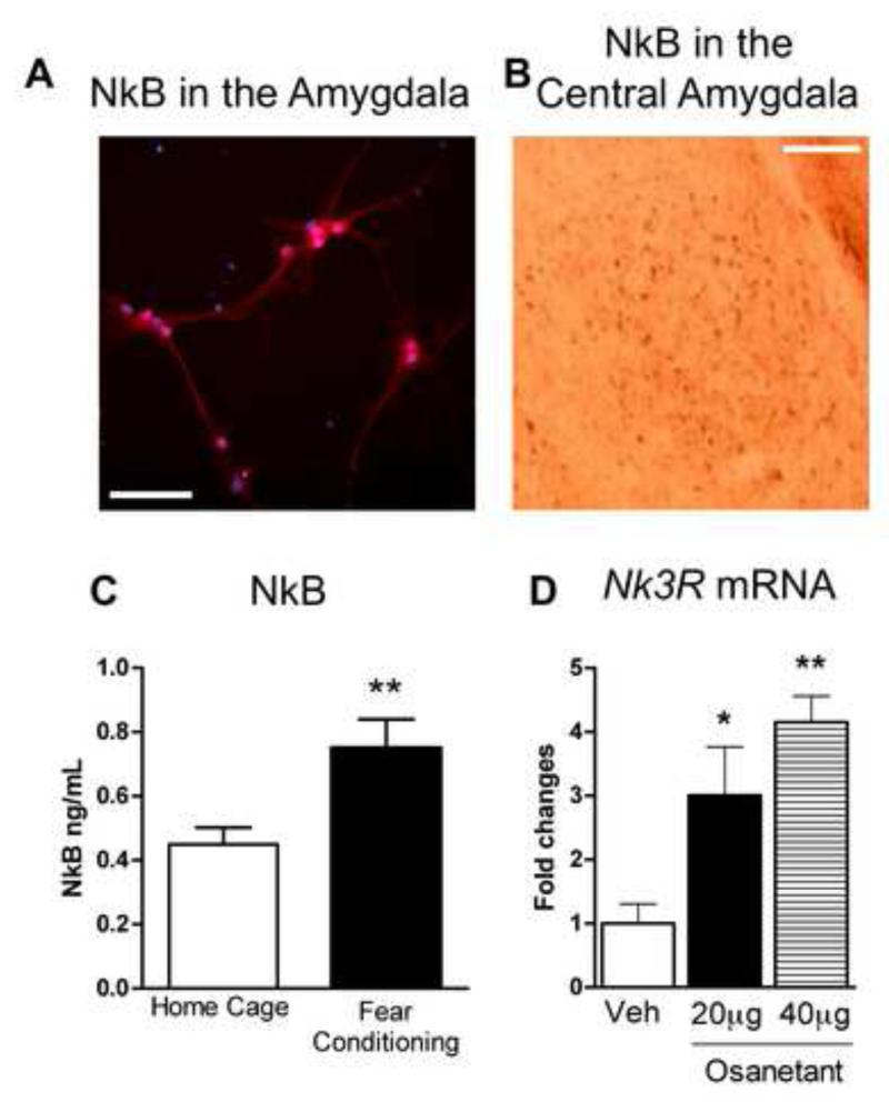
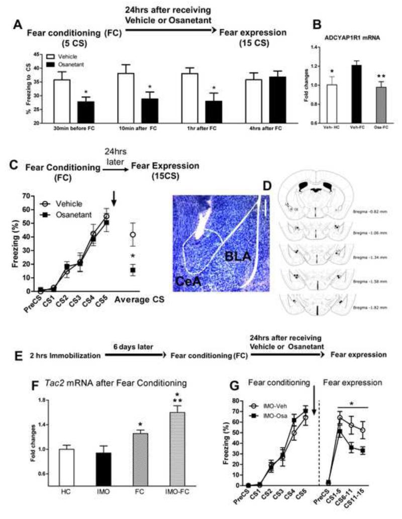
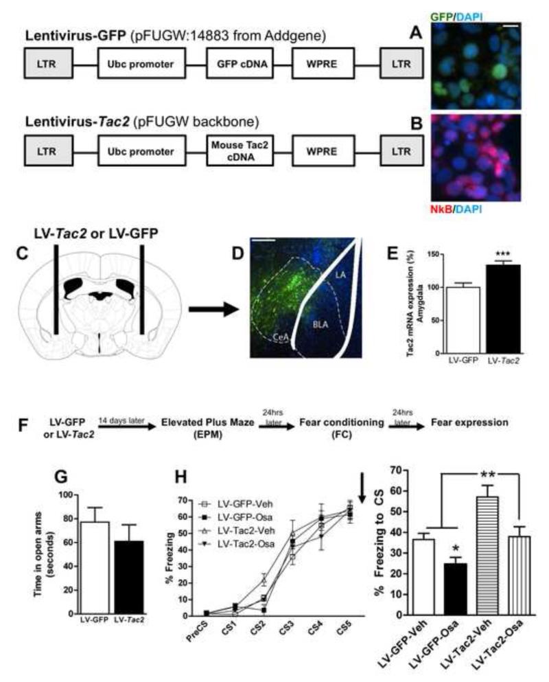
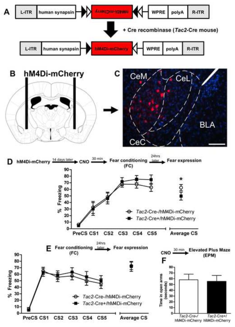
The centromedial amygdala (CeM), a subdivision of the central amygdala (CeA), is believed to be the main output station of the amygdala for fear expression. We provide evidence that the Tac2 gene, expressed by neurons specifically within the CeM, is required for modulating fear memories. Tac2 is colocalized with GAD65 and CaMKIIα but not with PKCd and Enk neurons in the CeM. Moreover, the Tac2 product, NkB, and its specific receptor, Nk3R, are also involved in the consolidation of fear memories. Increased Tac2 expression, through a stress-induced PTSD-like model, or following lentiviral CeA overexpression, are sufficient to enhance fear consolidation. This effect is blocked by the Nk3R antagonist osanetant. Concordantly, silencing of Tac2-expressing neurons in CeA with DREADDs impairs fear consolidation. Together, these studies further our understanding of the role of the Tac2 gene and CeM in fear processing and may provide approaches to intervention for fear-related disorders.


Figures

Figure 1
Figure 1. Differential regulation of Tac2 gene expression in the amygdala during cued-fear conditioning
A) With average linkage hierarchical clustering of a RNA microarray there is a differential gene regulation 30 minutes and 2 hours after auditory fear conditioning (FC) when compared to home cage group (no FC). N=4 per group. B) Tac2 mRNA levels are rapidly up-regulated in the amygdala during fear consolidation 30 minutes after fear conditioning. *P ≤0.05 vs HC and 2 hrs. N=7-8 per group. C) Tac2 up-regulation occurs when the conditioned stimulus (acoustic tone) and the unconditioned stimulus (electric footshock) are paired but not when they are unpaired. *P ≤0.05 vs HC and unpaired. N=11-15 per group. Mean + SEM is shown. D) Tac2 expression by radioactive in situ hybridization in the amygdala is restricted to the central amygdala (CeA) with highest expression in the CeM amygdala. Scale bar = 1 mm. See also Figure S1 and Table S1 and S2.
Figure 2
Figure 2. Tac2 is colocalized with glutamate decarboxylase 65 (GAD65) and calmodulin-dependent protein kinase II α (CAMKIIα) but is not colocalized with Protein Kinase C Delta nor Enkephalin-expressing neurons in the CeM
A and B) Tac2 mRNA expression in the CeA and BLA by non-radioactive fluorescent in situ hybridization (FISH). Scale bar = 100 μm. C) PKCd mRNA expression by FISH. Scale bar = 100 μm. D) Enk mRNA expression by FISH. Scale bar = 100 μm. E) Right, A and C merged showing different pattern of expression of Tac2 and PKCd in the CeA. Scale bar = 100 μm. Left, confocal image showing no colocalization of Tac2 and PKCd in the CeM. Scale bar = 15 μm. F) Right, B and D merged showing different pattern of expression of Tac2 and Enk. Scale bar = 100 μm. Left, confocal image showing no colocalization of Tac2 and Enk in the CeM. Scale bar = 15 μm. G) Confocal image showing colocalization of Tac2 mRNA expression and GAD65 peptide in the CeM. Scale bar = 15 μm. H) Confocal image showing colocalization of Tac2 mRNA expression and CaMKIIα peptide in the CeM. Scale bar = 15 μm. CeM = centro-medial amygdala, CeL = centro-lateral amygdala, CeC = centro-central amygdala, CeA = central amygdala, BLA = basolateral amygdala.
Figure 3
Figure 3. Fear conditioning, expression of Neurokinin B and Neurokinin 3 receptor in the amygdala
A) The Tac2 product, Neurokinin B (NkB) is detected by immunocytochemistry in mouse amygdala cell culture. NkB is highly expressed in the soma and in the dendrites. Red is NkB signal. Blue is neuronal nucleus, NeuN. Scale bar = 25 μm. B) Immunohistochemistry studies show high expression of NkB in the central amygdala (CeA). Scale bar = 125 μm. C) NkB is up-regulated at 2 hrs in the amygdala after fear conditioning. **P ≤0.01 vs Home Cage, N=6-8 per group. D) Amygdala cell culture with osanetant, a potent and specific neurokinin 3 (Nk3R) antagonist. Incubation with 20 μg and 40 μg of osanetant enhances Nk3R mRNA levels. This suggests that osanetant activates Nk3R and its downstream signaling in the amygdala. *P ≤0.05 vs Veh, **P ≤0.01 vs Veh, N=2 per group. Mean + SEM is shown.
Figure 4
Figure 4. A Nk3R antagonist impairs cued-fear memory consolidation when infused systemically, in the central amygdala and in a PTSD-like mouse model
A) Osanetant impairs cued-fear memory when given from 30 minutes to up to 1 hour after fear acquisition. The figure shows the time spent in freezing behavior during the fear expression test when the CS is presented. *P ≤.05 vs Veh. N=4-12 per group. B) Osanetant given intraperitoneally 30 minutes before fear conditioning impaired the enhancement of mRNA levels of the Pac1 receptor (Adcyap1r1), *P ≤0.05 vs Veh-FC; **P ≤0.01 vs Veh-FC. N= 9-12 per group. The PACAP-PAC1R pathway is associated with PTSD, fear conditioning and stress C) Osanetant bilaterally injected into the central amygdala immediately after fear conditioning causes impaired fear memory consolidation as shown by lower freezing in the cued-fear expression test *P ≤ 0.05, N= 3-9 mice per group. D) Histological verification of osanetant infusion sites. Left, toluidine blue staining showing an example of the tip of the cannula in the CeA. Scale bar = 250 μm. Right, the dots indicate the lowest point of the injector tip. E) Timeline of the experiment. F) Cued-fear conditioning enhances Tac2 levels 30 minutes after fear conditioning in naïve mice but more robustly in mice with a previous exposure to immobilization to a wooden board (IMO), a PTSD-like model. N=12-15 per group. *P ≤0.05 vs HC, **P ≤0.01 vs IMO. G) Osanetant was given immediately after FC and impaired fear memory consolidation in mice which had been previously exposed to a traumatic stress as shown by reduced freezing in the fear expression test, *P ≤0.05. N= 8 per group. Mean + or ± SEM is shown. Veh = vehicle; Osa = osanetant.
Figure 5
Figure 5. Tac2 overexpression in the central amygdala is sufficient to enhance fear memory consolidation and it is blocked by an Nk3R antagonist
A) The lentivirus GFP-FUGW induces GFP expression but not Neurokinin B (NkB) in Hek293 cells. Scale bar = 10 μm. B) The lentivirus Tac2-FUGW induces NkB expression in Hek293 cells. DAPI staining (blue) indicates the cellular nuclei. NkB staining (red) is contrasted with GFP fluorescence (green). C) Tac2-FUGW or GFP-FUGW were bilaterally infused in the central amygdala and mice were left undisturbed for 14 days. D-E) The lentivius Tac2-FUGW causes a 42% overexpression of Tac2 in the central amygdala. ***P ≤0.001 vs LV-GFP. N= 9-15 per group. Scale bar = 250 μm. F) Timeline of the experiment. G) Tac2 overexpression in the central amygdala does not alter anxiety-like behavior evaluated by the time spent in the open arms in the elevated plus maze. N= 9-15. H) Left, the lentiviruses GFP-FUGW and Tac2-FUGW causes no changes in fear conditioning. Osanetant or vehicle were given systemically immediately after fear acquisition. Right) Tac2 overexpression enhances fear memory consolidation (LV-Tac2-Veh) and osanetant impairs this effect (LV-Tac2-Osa). N= 3-8 per group. *P≤0.05 vs LV-GFP-Veh, **P ≤0.01 vs all other groups. Mean + or ± SEM is shown. See also Figure S5.
Figure 6
Figure 6. Inducible silencing of Tac2-expressing neurons in the CeA with Gi-DREADD decreases conditioned fear
A) Design of hM4Di-mCherry AAV and Tac2-cre mice. B and C) Tac2-Cre- or Tac2-Cre+ mice were infected with the hM4Di-mCherry AAV in the CeA. The Gi receptor was inserted only on the Tac2-Cre cells of Tac2-Cre+ mice, CeM = centro-medial amygdala, CeL = centro-lateral amygdala. Scale bar = 125 μm. D) CNO was given systemically 30 minutes prior to fear conditioning to Tac2-Cre-/hM4Di-mCherry and Tac2-Cre+/hM4Di-mCherry. Temporal silencing of the Tac2-expressing neurons in the Tac2-Cre+/hM4Di-mCherry group did not affect freezing during fear acquisition. However, when mice were tested the day after for fear expression, without CNO, Tac2-Cre+/hM4Di-mCherry mice showed less conditioned fear. *P ≤0.05 Tac2-Cre-/hM4Di-mCherry vs Tac2-Cre+/hM4Di-mCherry, N=10-11 per group. E) Tac2-Cre-/hM4Di-mCherry and Tac2-Cre+/hM4Di-mCherry mice were retrained to a different acoustic tone (CS) without receiving CNO. Both groups equally acquired fear learning and showed similar levels of fear memory consolidation. F) CNO given 30 minutes before the elevated plus maze showed no effect on Tac2-Cre-/hM4Di-mCherry nor Tac2-Cre+/hM4Di-mCherry in anxiety-like behavior. See also Figure S6.

References

    1. American Psychiatric Association . Diagnostic and Statistical Manual of Mental Disorders, Fourth Edition, Text Revision (DSM5) American Psychiatric Association; Washington, DC: 2013.
    1. Almli LM, Mercer KB, Kerley K, Feng H, Bradley B, Conneely KN, Ressler KJ. ADCYAP1R1 genotype associates with post-traumatic stress symptoms in highly traumatized African-American females. American journal of medical genetics Part B, Neuropsychiatric genetics: the official publication of the International Society of Psychiatric Genetics. 2013;162B:262–272. - PMC - PubMed
    1. Andero R, Brothers SP, Jovanovic T, Chen YT, Salah-Uddin H, Cameron M, Bannister TD, Almli L, Stevens JS, Bradley B, et al. Amygdala-dependent fear is regulated by Oprl1 in mice and humans with PTSD. Science translational medicine. 2013;5:188ra173. - PMC - PubMed
    1. Andero R, Heldt SA, Ye K, Liu X, Armario A, Ressler KJ. Effect of 7,8-dihydroxyflavone, a small-molecule TrkB agonist, on emotional learning. The American journal of psychiatry. 2011;168:163–172. - PMC - PubMed
    1. Andero R, Ressler KJ. Fear extinction and BDNF: translating animal models of PTSD to the clinic. Genes, brain, and behavior. 2012;11:503–512. - PMC - PubMed

Publication types

MeSH terms

Associated data