

Brain Regions Underlying Repetition and Auditory-Verbal Short-term Memory Deficits in Aphasia: Evidence from Voxel-based Lesion Symptom Mapping
- PMID: 24976669
- PMCID: PMC4070523
- DOI: 10.1080/02687038.2011.602391
Brain Regions Underlying Repetition and Auditory-Verbal Short-term Memory Deficits in Aphasia: Evidence from Voxel-based Lesion Symptom Mapping
Abstract
Background: A deficit in the ability to repeat auditory-verbal information is common among individuals with aphasia. The neural basis of this deficit has traditionally been attributed to the disconnection of left posterior and anterior language regions via damage to a white matter pathway, the arcuate fasciculus. However, a number of lesion and imaging studies have called this notion into question.
Aims: The goal of this study was to identify the neural correlates of repetition and a related process, auditory-verbal short-term memory (AVSTM). Both repetition and AVSTM involve common elements such as auditory and phonological analysis and translation to speech output processes. Based on previous studies, we predicted that both repetition and AVSTM would be most dependent on posterior language regions in left temporo-parietal cortex.
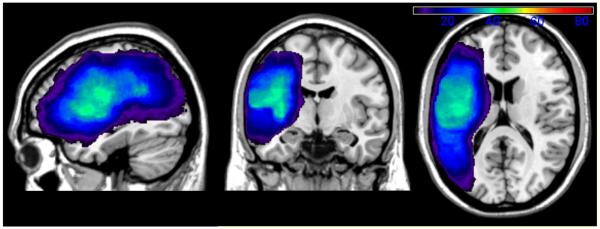
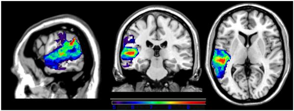
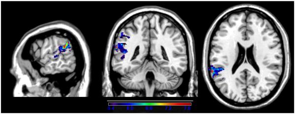
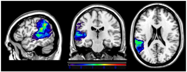
Methods & procedures: We tested 84 individuals with left hemisphere lesions due to stroke on an experimental battery of repetition and AVSTM tasks. Participants were tested on word, pseudoword, and number-word repetition, as well as digit and word span tasks. Brain correlates of these processes were identified using a statistical, lesion analysis approach known as voxel-based lesion symptom mapping (VLSM). VLSM allows for a voxel-by-voxel analysis of brain areas most critical to performance on a given task, including both grey and white matter regions.
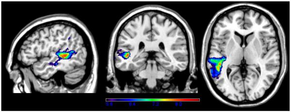
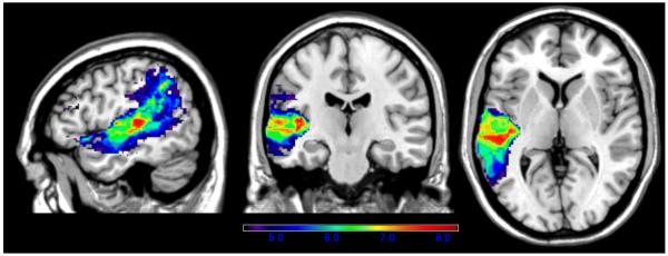
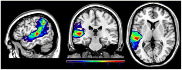
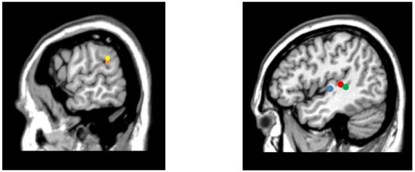
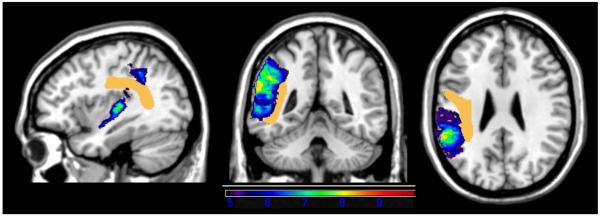
Outcomes & results: The VLSM analyses showed that left posterior temporo-parietal cortex, not the arcuate fasciculus, was most critical for repetition as well as for AVSTM. The location of maximal foci, defined as the voxels with the highest t values, varied somewhat among measures: Word and pseudoword repetition had maximal foci in the left posterior superior temporal gyrus, on the border with inferior parietal cortex, while word and digit span, as well as number-word repetition, were centered on the border between the middle temporal and superior temporal gyri and the underlying white matter.
Conclusions: Findings from the current study show that 1) repetition is most critically mediated by cortical regions in left posterior temporo-parietal cortex; 2) repetition and AVSTM are mediated by partially overlapping networks; and 3) repetition and AVSTM deficits can be observed in different types of aphasia, depending on the site and extent of the brain injury. These data have implications for the prognosis of chronic repetition and AVSTM deficits in individuals with aphasia when lesions involve critical regions in left temporo-parietal cortex.
Keywords: aphasia; conduction aphasia; parietal cortex; repetition; short-term memory; temporal cortex.
Figures
References
-
- Anderson JM, Gilmore R, Roper S, Crosson B, et al. Conduction aphasia and the arcuate fasciculus: A reexamination of the Wernicke-Geschwind model. Brain and Language. 1999;70:1–12. - PubMed
-
- Awh E, Smith E, Jonides J. Human rehearsal processes and the frontal lobes: PET evidence. Annals of the New York Academy of Sciences. 1995;769:97–117. - PubMed
-
- Axer H, Keyserlingk A, Berks G, Keyserlingk D. Supra- and infrafsylvian conduction aphasia. Brain and Language. 2001;76:317–331. - PubMed
-
- Baddeley A, Hitch G. Developments in the concept of working memory. Neuropsychology. 1994;8:485–493.
Grants and funding
LinkOut - more resources
Full Text Sources