

Endoscopic ultrasound in the diagnosis of pancreatic intraductal papillary mucinous neoplasms
- PMID: 24976716
- PMCID: PMC4069307
- DOI: 10.3748/wjg.v20.i24.7785
Endoscopic ultrasound in the diagnosis of pancreatic intraductal papillary mucinous neoplasms
Abstract
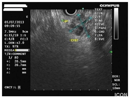
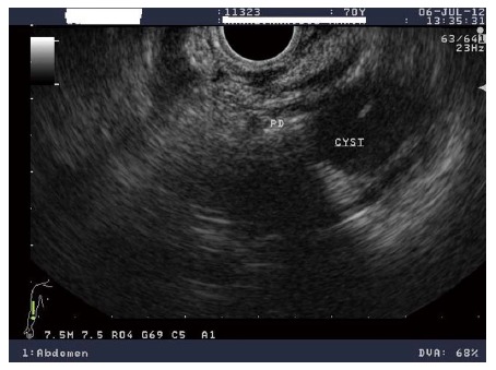
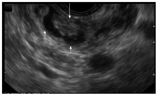
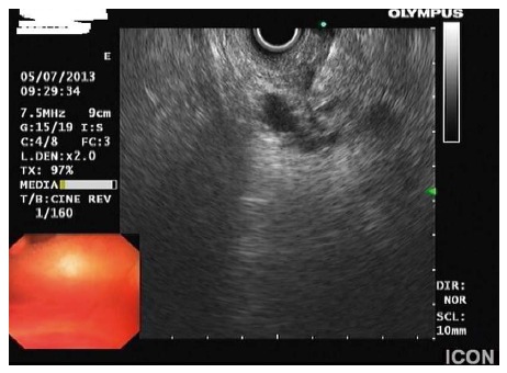
Pancreatic cystic lesions are increasingly recognised due to the widespread use of different imaging modalities. Intraductal papillary mucinous neoplasms (IPMNs) of the pancreas represent a common, but also heterogeneous group of cystic tumors with a significant malignant potential. These neoplasms must be differentiated from other cystic tumors and properly classified into their different types, main-duct IPMNs vs branch-duct IPMNs. These types have a different malignant potential and therefore, different treatment strategies need to be implemented. Endoscopic ultrasound (EUS) offers the highest resolution of the pancreas and can aid in the differential diagnosis, classification and differentiation between benign and malignant tumors. The addition of EUS fine-needle aspiration can supply further information by obtaining fluid for cytology, measurement of tumor markers and perhaps DNA analysis. Novel techniques, such as the use of contrast and sophisticated equipment, like intraductal probes can provide information regarding malignant features and extent of these neoplasms. Thus, EUS is a valuable tool in the diagnosis and appropriate management of these tumors.
Keywords: Endoscopic ultrasound; Pancreatic intraductal papillary mucinous neoplasms.
Figures
Similar articles
-
The role of endoscopic ultrasound in the management of intraductal papillary mucinous neoplasms.Minerva Med. 2014 Oct;105(5):413-21. Epub 2014 Jul 7. Minerva Med. 2014. PMID: 25000220 Review.
-
Management of branch-duct intraductal papillary mucinous neoplasms: a large single-center study to assess predictors of malignancy and long-term outcomes.Gastrointest Endosc. 2016 Sep;84(3):436-45. doi: 10.1016/j.gie.2016.02.008. Epub 2016 Feb 18. Gastrointest Endosc. 2016. PMID: 26905937
-
Imaging and Cytopathological Criteria Indicating Malignancy in Mucin-Producing Pancreatic Neoplasms: A Series of 68 Histopathologically Confirmed Cases.Pancreas. 2018 Nov/Dec;47(10):1283-1289. doi: 10.1097/MPA.0000000000001182. Pancreas. 2018. PMID: 30308535
-
Endoscopic ultrasonographic findings predict the risk of carcinoma in branch duct intraductal papillary mucinous neoplasms of the pancreas.Pancreatology. 2012 Mar-Apr;12(2):141-5. doi: 10.1016/j.pan.2011.12.008. Epub 2012 Jan 14. Pancreatology. 2012. PMID: 22487524
-
The role of endoscopic ultrasound in the management of intraductal papillary mucinous neoplasms: a systematic update.Minerva Med. 2016 Dec;107(6):370-380. Epub 2016 Sep 14. Minerva Med. 2016. PMID: 27627636 Review.
Cited by
-
Basic Principles and Role of Endoscopic Ultrasound in Diagnosis and Differentiation of Pancreatic Cancer from Other Pancreatic Lesions: A Comprehensive Review of Endoscopic Ultrasound for Pancreatic Cancer.J Clin Med. 2024 Apr 28;13(9):2599. doi: 10.3390/jcm13092599. J Clin Med. 2024. PMID: 38731128 Free PMC article. Review.
-
The effectiveness of endoscopic ultrasonography findings to distinguish benign and malignant intraductal papillary mucinous neoplasm.Surg Endosc. 2023 Jun;37(6):4681-4688. doi: 10.1007/s00464-022-09752-3. Epub 2023 Mar 7. Surg Endosc. 2023. PMID: 36881188 Free PMC article. Clinical Trial.
-
Multimodality imaging studies of intraductal tubulopapillary neoplasms of the pancreas.Diagn Interv Radiol. 2019 Jul;25(4):251-256. doi: 10.5152/dir.2019.18215. Diagn Interv Radiol. 2019. PMID: 31147310 Free PMC article.
-
Advantages of deep learning reconstruction algorithm in ultra-high-resolution CT for the diagnosis of pancreatic cystic neoplasm.Jpn J Radiol. 2025 May 30. doi: 10.1007/s11604-025-01804-7. Online ahead of print. Jpn J Radiol. 2025. PMID: 40445272
-
Pancreatic cystic neoplasms: Review of current knowledge, diagnostic challenges, and management options.J Carcinog. 2015 Mar 14;14:3. doi: 10.4103/1477-3163.153285. eCollection 2015. J Carcinog. 2015. PMID: 25821410 Free PMC article. Review.
References
-
- Ohashi K, Murakami Y, Takekoshi T. Four cases of mucin producing cancer of the pancreas on specific findings of the papilla of Vater. Prof Dig Endosc. 1982;20:348–351.
-
- Kloppel G, Solcia E, Longnecker DS, Capella C, Sobin LH. World Health Organization international histologic typing of tumors of the exocrine pancreas. In: Classifications of tumors., editor. Berlin, Germany: Springer; 1996. pp. 11–20.
-
- Brugge WR, Lauwers GY, Sahani D, Fernandez-del Castillo C, Warshaw AL. Cystic neoplasms of the pancreas. N Engl J Med. 2004;351:1218–1226. - PubMed
-
- Yoon WJ, Lee JK, Lee KH, Ryu JK, Kim YT, Yoon YB. Cystic neoplasms of the exocrine pancreas: an update of a nationwide survey in Korea. Pancreas. 2008;37:254–258. - PubMed
Publication types
MeSH terms
LinkOut - more resources
Full Text Sources
Other Literature Sources
Medical
