

Molecular confocal laser endomicroscopy: a novel technique for in vivo cellular characterization of gastrointestinal lesions
- PMID: 24976717
- PMCID: PMC4069308
- DOI: 10.3748/wjg.v20.i24.7794
Molecular confocal laser endomicroscopy: a novel technique for in vivo cellular characterization of gastrointestinal lesions
Abstract
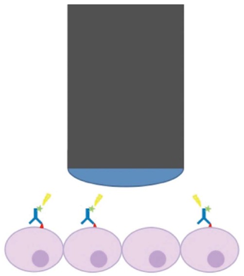
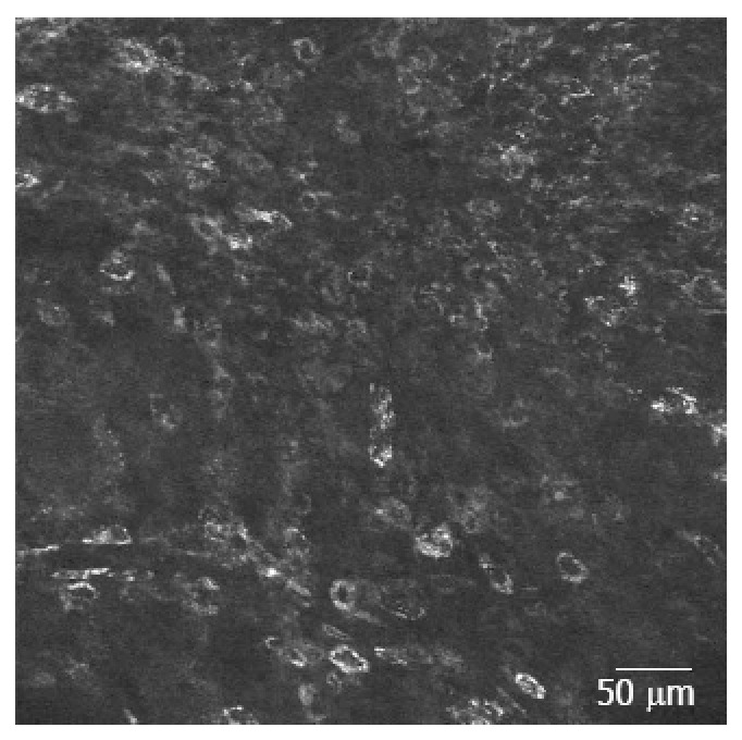
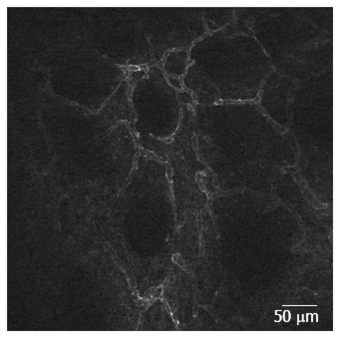
While flexible endoscopy is essential for macroscopic evaluation, confocal laser endomicroscopy (CLE) has recently emerged as an endoscopic method enabling visualization at a cellular level. Two systems are currently available, one based on miniprobes that can be inserted via a conventional endoscope or via a needle guided by endoscopic ultrasound. The second system has a confocal microscope integrated into the distal part of an endoscope. By adding molecular probes like fluorescein conjugated antibodies or fluorescent peptides to this procedure (either topically or systemically administered during on-going endoscopy), a novel world of molecular evaluation opens up. The method of molecular CLE could potentially be used for estimating the expression of important receptors in carcinomas, subsequently resulting in immediate individualization of treatment regimens, but also for improving the diagnostic accuracy of endoscopic procedures by identifying otherwise invisible mucosal lesions. Furthermore, studies have shown that fluorescein labelled drugs can be used to estimate the affinity of the drug to a target organ, which probably can be correlated to the efficacy of the drug. However, several of the studies in this research field have been conducted in animal facilities or in vitro, while only a limited number of trials have actually been carried out in vivo. Therefore, safety issues still needs further evaluations. This review will present an overview of the implications and pitfalls, as well as future challenges of molecular CLE in gastrointestinal diseases.
Keywords: Barrett’s esophagus; Colorectal carcinoma; Confocal laser endomicroscopy; Endoscopy imaging; Gastric carcinoma; Inflammatory bowel disease.
Figures
References
-
- Hirschowitz BI, Curtiss LE, Peters CW, Pollard HM. Demonstration of a new gastroscope, the fiberscope. Gastroenterology. 1958;35:50; discussion 51–53. - PubMed
-
- Goetz M. Real-time histology in colonoscopy. Gastroenterol Clin North Am. 2013;42:567–575. - PubMed
-
- Yarza P, Spröer C, Swiderski J, Mrotzek N, Spring S, Tindall BJ, Gronow S, Pukall R, Klenk HP, Lang E, et al. Sequencing orphan species initiative (SOS): Filling the gaps in the 16S rRNA gene sequence database for all species with validly published names. Syst Appl Microbiol. 2013;36:69–73. - PubMed
-
- Qumseya BJ, Wang H, Badie N, Uzomba RN, Parasa S, White DL, Wolfsen H, Sharma P, Wallace MB. Advanced imaging technologies increase detection of dysplasia and neoplasia in patients with Barrett’s esophagus: a meta-analysis and systematic review. Clin Gastroenterol Hepatol. 2013;11:1562–70.e1-2. - PMC - PubMed
-
- Shahid MW, Buchner AM, Heckman MG, Krishna M, Raimondo M, Woodward T, Wallace MB. Diagnostic accuracy of probe-based confocal laser endomicroscopy and narrow band imaging for small colorectal polyps: a feasibility study. Am J Gastroenterol. 2012;107:231–239. - PubMed
Publication types
MeSH terms
Substances
LinkOut - more resources
Full Text Sources
Other Literature Sources
