Review
. 2014 Jun 28;20(24):7864-77.
doi: 10.3748/wjg.v20.i24.7864.

Imaging diagnosis of pancreatic cancer: a state-of-the-art review

Affiliations
Review

Imaging diagnosis of pancreatic cancer: a state-of-the-art review

Eun Sun Lee et al. World J Gastroenterol. .

Abstract

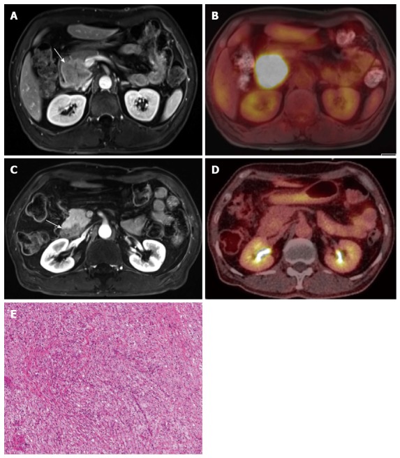
Pancreatic cancer (PC) remains one of the deadliest cancers worldwide, and has a poor, five-year survival rate of 5%. Although complete surgical resection is the only curative therapy for pancreatic cancer, less than 20% of newly-diagnosed patients undergo surgical resection with a curative intent. Due to the lack of early symptoms and the tendency of pancreatic adenocarcinoma to invade adjacent structures or to metastasize at an early stage, many patients with pancreatic cancer already have advanced disease at the time of their diagnosis and, therefore, there is a high mortality rate. To improve the patient survival rate, early detection of PC is critical. The diagnosis of PC relies on computed tomography (CT) and/or magnetic resonance imaging (MRI) with magnetic resonance cholangiopancreatography (MRCP), or biopsy or fine-needle aspiration using endoscopic ultrasound (EUS). Although multi-detector row computed tomography currently has a major role in the evaluation of PC, MRI with MRCP facilitates better detection of tumors at an early stage by allowing a comprehensive analysis of the morphological changes of the pancreas parenchyma and pancreatic duct. The diagnosis could be improved using positron emission tomography techniques in special conditions in which CT and EUS are not completely diagnostic. It is essential for clinicians to understand the advantages and disadvantages of the various pancreatic imaging modalities in order to be able to make optimal treatment and management decisions. Our study investigates the current role and innovative techniques of pancreatic imaging focused on the detection of pancreatic cancer.

Keywords: Endoscopic ultrasound-guided fine needle aspiration; Magnetic resonance imaging; Multi-detector computed tomography; Pancreatic neoplasms; Positron-emission tomography; Ultrasonography.


Figures

Figure 1
Figure 1
A 58-year-old, male patient with pancreatic body cancer with typical imaging findings. A, B: Endoscopic ultrasonography shows an approximately 3-cm, hypoechoic mass (arrows) in the pancreatic body with distal pancreatic duct dilatation; C, D: The mass (arrow) shows hypointensity on portal-venous-phase, contrast enhanced MR and hypermetabolism on PET/CT, respectively. MR: Magnetic resonance; PET: Positron emission tomography; CT: Computed tomography.
Figure 2
Figure 2
A 73-year-old male with pathologically-proven pancreatic head cancer. A: Approximately 3 cm low attenuating mass (arrow) is noted at the pancreatic head on the CT scan; B: In pre-contrast T1-weighted gradient echo sequence of MR, this mass (arrow) shows lower signal intensity, compared to the normal pancreatic parenchyma; C: After contrast media administration, the pancreatic head cancer (arrow) has poor enhancement; D, E: DWI with 1000 of b-value and ADC map reveal the diffusion restriction of the pancreatic head cancer (arrow). CT: Computed tomography; MR: Magnetic resonance; DWI: Diffusion weighted imaging; ADC: Apparent diffusion coefficient.
Figure 3
Figure 3
A 64-year-old male with biopsy-proven pancreatic adenocarcinoma with liver metastasis. A, B: On MDCT, the pancreatic tail mass (arrow) shows isoattenuation, causing distal parenchyma atrophy; C: On pre-contrast, T1–weighted, gradient-echo sequence MRI, the pancreatic tail mass (arrow) is clearly depicted, as well as the liver metastasis, owing to the increased soft-tissue contrast of MR compared with that of CT. MRI: Magnetic resonance imaging; MDCT: Multi-detector computed tomography.
Figure 4
Figure 4
Post-process of multi-detector computed tomography for pancreatic cancer. A: In the axial CT scan, an ill-defined pancreatic body cancer invading the celiac axis is identified; B: On the curved MPR along the pancreatic duct, the relationship between the pancreatic duct and the cancer can be more easily understood; C, D: The extent and degree of major vascular involvement caused by the pancreatic cancer can be comprehensively assessed using MPR and the maximum intensity projections. MDCT: Multi-detector computed tomography; MPR: Multiplanar reformations.
Figure 5
Figure 5
Treatment monitoring of pancreatic cancer using positron emission tomography/magnetic resonance. A, B: A 5-cm mass of biopsy-proven, adenosquamous carcinoma (arrow) in the pancreatic head, as seen due to the strong FDG uptake; C, D: The mass (arrow) shows a marked decrease in size and glucose metabolism (from 22.0 to 3.8 of mSUV) after six cycles of neoadjuvant concurrent chemoradiation treatment. The specimen obtained during the following surgery revealed complete remission; E: All tumor cells are replaced by a foamy histiocytes collection of cholesterol clefts and multinucleated giant cells. PET: Positron emission tomography; MR: Magnetic resonance; FDG: Fluorodeoxyglucose; mSUV: Maximum standardized uptake value.

Similar articles

Cited by

References

    1. Hariharan D, Saied A, Kocher HM. Analysis of mortality rates for pancreatic cancer across the world. HPB (Oxford) 2008;10:58–62. - PMC - PubMed
    1. Hidalgo M. Pancreatic cancer. N Engl J Med. 2010;362:1605–1617. - PubMed
    1. Siegel R, Naishadham D, Jemal A. Cancer statistics, 2013. CA Cancer J Clin. 2013;63:11–30. - PubMed
    1. Li D, Xie K, Wolff R, Abbruzzese JL. Pancreatic cancer. Lancet. 2004;363:1049–1057. - PubMed
    1. Jemal A, Siegel R, Ward E, Hao Y, Xu J, Murray T, Thun MJ. Cancer statistics, 2008. CA Cancer J Clin. 2008;58:71–96. - PubMed

MeSH terms