Review
. 2014 Feb;22(1):2-12.
doi: 10.1179/2042618613Y.0000000060.

Temporomandibular disorders. Part 1: anatomy and examination/diagnosis

Affiliations
Review

Temporomandibular disorders. Part 1: anatomy and examination/diagnosis

Stephen M Shaffer et al. J Man Manip Ther. 2014 Feb.

Abstract

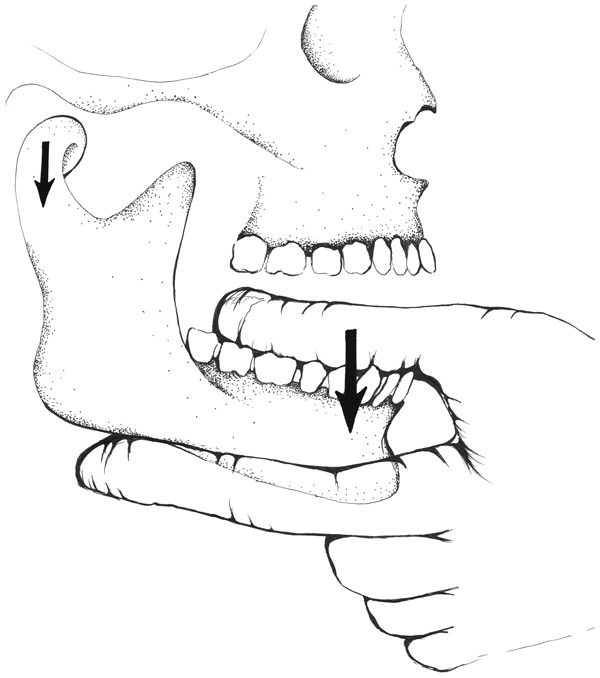
Temporomandibular disorders (TMD) are a heterogeneous group of diagnoses affecting the temporomandibular joint (TMJ) and surrounding tissues. A variety of methods for evaluating and managing TMD have been proposed within the physical therapy profession but these sources are not peer-reviewed and lack updates from scientific literature. The dental profession has provided peer-reviewed sources that lack thoroughness with respect to the neuromusculoskeletal techniques utilized by physical therapists. The subsequent void creates the need for a thorough, research informed, and peer-reviewed source regarding TMD evaluation and management for physical therapists. This paper is the first part in a two-part series that seeks to fill the current void by providing a brief but comprehensive outline for clinicians seeking to provide services for patients with TMD. Part one focuses on anatomy and pathology, arthro- and osteokinematics, epidemiology, history taking, and physical examination as they relate to TMD. An appreciation of the anatomical and mechanical features associated with the TMJ can serve as a foundation for understanding a patient's clinical presentation. Performance of a thorough patient history and clinical examination can guide the clinician toward an improved diagnostic process.

Keywords: Diagnosis; Physical examination; Review; Temporomandibular joint disorders.


Figures

Figure 1
Figure 1
Superficial view of temporomandibular joint (TMJ) anatomy. (a) temporalis muscle, (b) temporomandibular ligament, (c) lateral pterygoid muscle, and (d) masseter muscle. © Jennifer Lenox.
Figure 2
Figure 2
Deep view of temporomandibular joint (TMJ) anatomy. (a) temporalis muscle, (b) temporalis bone, (c) mandibular condyle, (d) lateral pterygoid muscle, (e) medial pterygoid muscle, and (f) intraarticular disk. © Jennifer Lenox.
Figure 3
Figure 3
Temporomandibular joint (TMJ) arthrokinematics during mouth opening. (a) discotemporal space, (b) intraarticular disk, (c) discomandibular space, (d) mandibular condyle, (e) posterior joint capsule, (f) temporalis bone, (g) upper and lower heads of the lateral pterygoid muscle, arrow 1a: posterior rolling of mandibular condyle, arrow 1b: anterior-caudal translation of mandibular condyle, and arrow 2: anterior-caudal translation of disk and mandibular condyle. Discomandibular rotation involves a combination of arthrokinematic posterior rolling (arrow 1a) and anterior translation (arrow 1b). Discotemporal anterior translation occurs as both the intraarticular disk and mandibular condyle simultaneously glide in an anterior-caudal direction along the inferior slope of the temporalis bone (arrow 2). © Jennifer Lenox.
Figure 4
Figure 4
Boley gage. When measuring mouth opening, the upper notch facing left is stabilized on the inferior aspect of the upper incisors. Once stabilized, the portion of the Boley gage with the lower notch facing left is slid down the measurement scale until the lower notch contacts the superior aspect of the lower incisors. The instrument’s scale is then read and the opening range obtained.
Figure 5
Figure 5
TheraBite range-of-motion (ROM) scale. When measuring mouth opening, the notch at the lower left portion of the scale is stabilized on the superior aspect of the lower incisors. Once stabilized, the instrument is rotated up toward the inferior aspect of upper incisors until contact is made between the scale and the upper incisors. The instrument’s scale is then read and the opening range obtained.
Figure 6
Figure 6
Hand placement for intraoral passive accessory motion testing. Large arrow: distraction force placed through the ipsilateral lower molars and premolars by the first digit while the second digit provides a counterforce on the inferior aspect of the ipsilateral mandibular body. Utilizing the third digit to provide additional counterforce on the inferior aspect of the contralateral mandibular body may maximize patient comfort. Small arrow: direction of the distraction movement of the mandibular condyle at the temporomandibular joint (TMJ). © Jennifer Lenox.

Similar articles

Cited by

References

    1. McNeill C. History and evolution of TMD concepts. Oral Surg Oral Med Oral Pathol Oral Radiol Endod. 1997;83:51–60. - PubMed
    1. Griffiths RH. Report of the president’s conference on the examination, diagnosis, and management of temporomandibular disorders. J Am Dent Assoc. 1983;106:75–7. - PubMed
    1. Winkel D, Aufdemkampe G, Matthijs O, Meijer OG, Phelps V.Temporomandibular joint. Diagnosis and treatment of the spine: nonoperative orthopaedic medicine and manual therapy Gaithersburg: Aspen Publishers; 1996. p. 715–68.
    1. Widmer CG.Evaluation of temporomandibular disorders In: Krause SL, editor. TMJ disorders, management of the craniomandibular complexNew York, NY, USA: Churchill Livingstone; 1988. p. 79–112.
    1. Kaltenborn FM, Evjenth O, Baldauf Kaltenborn T, Morgan D, Vollowitz E. 2003. Manual mobilization of the joints: volume 2, the spine, 4th edn. Oslo.

LinkOut - more resources