Review
. 2014 Feb;22(1):13-23.
doi: 10.1179/2042618613Y.0000000061.

Temporomandibular disorders. Part 2: conservative management

Affiliations
Review

Temporomandibular disorders. Part 2: conservative management

Stephen M Shaffer et al. J Man Manip Ther. 2014 Feb.

Abstract

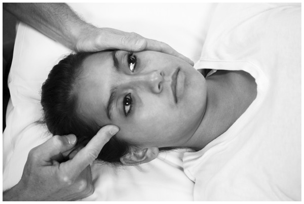
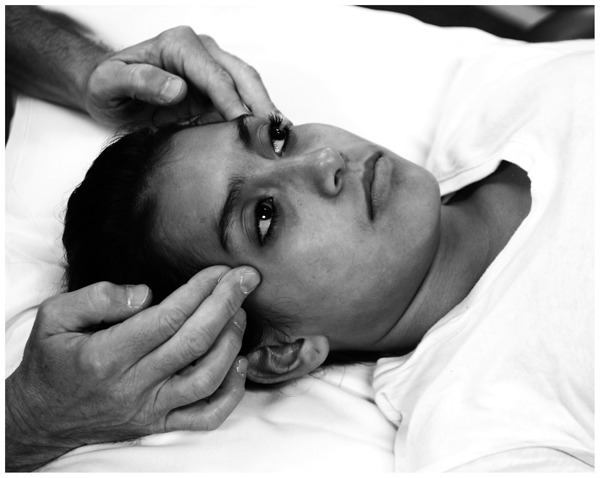
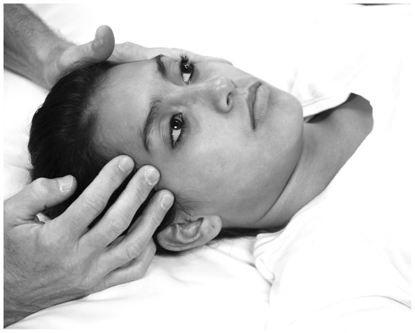
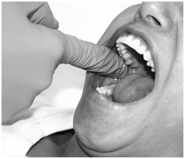
Appropriate management of temporomandibular disorders (TMD) requires an understanding of the underlying dysfunction associated with the temporomandibular joint (TMJ) and surrounding structures. A comprehensive examination process, as described in part 1 of this series, can reveal underlying clinical findings that assist in the delivery of comprehensive physical therapy services for patients with TMD. Part 2 of this series focuses on management strategies for TMD. Physical therapy is the preferred conservative management approach for TMD. Physical therapists are professionally well-positioned to step into the void and provide clinical services for patients with TMD. Clinicians should utilize examination findings to design rehabilitation programs that focus on addressing patient-specific impairments. Potentially appropriate plan of care components include joint and soft tissue mobilization, trigger point dry needling, friction massage, therapeutic exercise, patient education, modalities, and outside referral. Management options should address both symptom reduction and oral function. Satisfactory results can often be achieved when management focuses on patient-specific clinical variables.

Keywords: Conservative management; Review; Temporomandibular joint disorders.


Figures

Figure 1
Figure 1
Distraction of the temporomandibular joint (TMJ); large arrow: distraction force placed through the ipsilateral lower molars and premolars with the first digit while the second and third digits provide a counterforce on the inferior aspect of the ipsilateral and contralateral mandibular bodies, respectively; medium arrow: posteriorly directed stabilization force applied through the ipsilateral aspect of the patient’s forehead; small arrow: the examiner palpates the joint line to assess for movement of the mandibular condyle.
Figure 2
Figure 2
Anterior glide of the temporomandibular joint (TMJ); large arrow: anterior glide force with mild caudal bias placed through the mandible via gripping the ipsilateral lower molars and premolars with the first digit while the second and third digits provide a counterforce on the inferior aspect of the ipsilateral and contralateral mandibular bodies, respectively; medium arrow: posteriorly directed stabilization force applied through the ipsilateral aspect of the patient’s forehead; small arrow: the examiner palpates the joint line to assess for movement of the mandibular condyle.
Figure 3
Figure 3
Anterior glide of the temporomandibular joint (TMJ) with pre-positioned mouth opening; large arrow: anterior-inferior glide force placed through the mandible via gripping the ipsilateral lower molars and premolars with the first digit while the second and third digits provide a counterforce on the inferior aspect of the ipsilateral and contralateral mandibular bodies, respectively; medium arrow: posteriorly directed stabilization force applied through the ipsilateral aspect of the patient’s forehead; small arrow: the examiner palpates the joint line to assess for movement of the mandibular condyle.
Figure 4
Figure 4
Medial/lateral glide of the temporomandibular joint (TMJ); large arrow: medial glide force placed through the mandibular condyle and/or mandibular ramus; small arrow: the contralateral hand provides a stabilizing force either through the contralateral zygomatic arch of the temporal bone and/or the contralateral mandibular condyle, depending on the patient’s experience.
Figure 5
Figure 5
Caudal-anterior-medial (CAM) glide of the temporomandibular joint (TMJ); large arrow: combined caudal, anterior, and medial glide force placed through the mandibular condyle and/or mandibular ramus; small arrow: the contralateral hand provides a stabilizing force either through the contralateral zygomatic arch of the temporal bone and/or the contralateral mandibular condyle, depending on the patient’s experience.
Figure 6
Figure 6
Caudal-anterior-medial (CAM) glide of the temporomandibular joint (TMJ) with pre-positioned mouth opening; large arrow: medial glide force placed through the mandibular condyle and/or mandibular ramus; small arrow: the contralateral hand provides a stabilizing force either through the contralateral zygomatic arch of the temporal bone and/or the contralateral mandibular condyle, depending on the patient’s experience.
Figure 7
Figure 7
Self-mobilization of the temporomandibular joint (TMJ); large arrow: medial glide force placed through the mandibular condyle and/or mandibular ramus; small arrow: the contralateral hand provides a stabilizing force either through the contralateral zygomatic arch of the temporal bone and/or the contralateral mandibular condyle, depending on the patient’s experience.
Figure 8
Figure 8
Self-mobilization of the temporomandibular joint (TMJ) with pre-positioned mouth opening; large arrow: medial glide force placed through the mandibular condyle and/or mandibular ramus; small arrow: the contralateral hand provides a stabilizing force either through the contralateral zygomatic arch of the temporal bone and/or the contralateral mandibular condyle, depending on the patient’s experience.
Figure 9
Figure 9
Soft tissue mobilization of the temporalis muscle utilizing one digit for contact and one hand for contralateral stabilization. Palpation of an MTrP or the general muscle belly by the tip of one digit (second digit shown) to apply soft tissue mobilization to the temporalis muscle. Switching between different digits can be helpful in prolonging technique application time before the onset of fatigue. Note that a contralateral hand provides a counterforce to stabilize the head and prevent inadvertent head motion. In this instance, the therapist begins at the anterior margin of the muscle and moves posteriorly while focusing on areas of the muscle that require treatment.
Figure 10
Figure 10
Bilateral soft tissue mobilization of the temporalis muscle utilizing reinforced digits for contact. Palpation of an MTrP or the general muscle belly by the tips of the second digits with reinforcement by the third digits to apply soft tissue mobilization to the temporalis muscles. Note that both hands provide simultaneous treatment and therefore serve as both the treating and stabilizing forces. In this instance, the therapist begins at the anterior margin of the muscles and moves posteriorly.
Figure 11
Figure 11
Soft tissue mobilization of the temporalis muscle utilizing three digits for contact and one hand for contralateral stabilization. Palpation of the muscle belly by the tips of three digits to apply soft tissue mobilization to the right temporalis muscle. Switching pressure and emphasis to different digits can be helpful in prolonging technique application time before the onset of fatigue. Note that the contralateral hand provides a counterforce to stabilize the head and prevent inadvertent head motion. In this instance, the therapist begins at the anterior margin of the muscle and moves posteriorly.
Figure 12
Figure 12
Soft tissue mobilization of the medial pterygoid muscle. Palpation of the muscle belly by the tip of the second digit to apply soft tissue mobilization to the medial pterygoid muscle. For treatment purposes, having the patient relax so that the mouth is not opened widely can be advantageous. The open mouth position is utilized here for visualization purposes.

Similar articles

Cited by

References

    1. Young B, Walker MJ, Strunce J, Boyles R. A combined treatment approach emphasizing impairment-based manual physical therapy for plantar heel pain: a case series. J Orthop Sports Phys Ther. 2004;34:725–33. - PubMed
    1. Furto ES, Cleland JA, Whitman JM, Olson KA. Manual physical therapy interventions and exercise for patients with temporomandibular disorders. Cranio. 2006;24:283–91. - PubMed
    1. Walker MJ, Boyles RE, Young BA, Strunce JB, Garber MB, Whitman JM, et al. The effectiveness of manual physical therapy and exercise for mechanical neck pain: a randomized clinical trial. Spine. 2008;33:2371–8. - PubMed
    1. List T, Axelsson S. Management of TMD: evidence from systematic reviews and meta-analyses. J Oral Rehabil. 2010;37:430–51. - PubMed
    1. Bialosky JE, Bishop MD, Price DD, Robinson ME, George SZ. The mechanisms of manual therapy in the treatment of musculoskeletal pain: a comprehensive model. Man Ther. 2009;14:531–8. - PMC - PubMed

LinkOut - more resources