

Value of three dimensional power Doppler ultrasound in prediction of endometrial carcinoma in patients with postmenopausal bleeding
- PMID: 24976771
- PMCID: PMC4072554
- DOI: 10.5152/jtgga.2014.07355
Value of three dimensional power Doppler ultrasound in prediction of endometrial carcinoma in patients with postmenopausal bleeding
Abstract
Objective: To determine whether endometrial volume or power Doppler indices measured by 3-dimensional (3D) ultrasound imaging can discriminate between benign and malignant endometrium in women with postmenopausal bleeding and endometrial thickness ≥5 mm.
Material and methods: The current diagnostic accuracy study was conducted at Ain Shams University Maternity Hospital. Eighty-four patients with postmenopausal bleeding and endometrial thickness ≥5 mm underwent 3D power Doppler ultrasound examination of the corpus uteri. The endometrial volume was calculated, along with the vascularization index (VI), flow index (FI), and vascularization flow index (VFI) in the endometrium. The gold standard was the histopathological diagnosis of the endometrium.
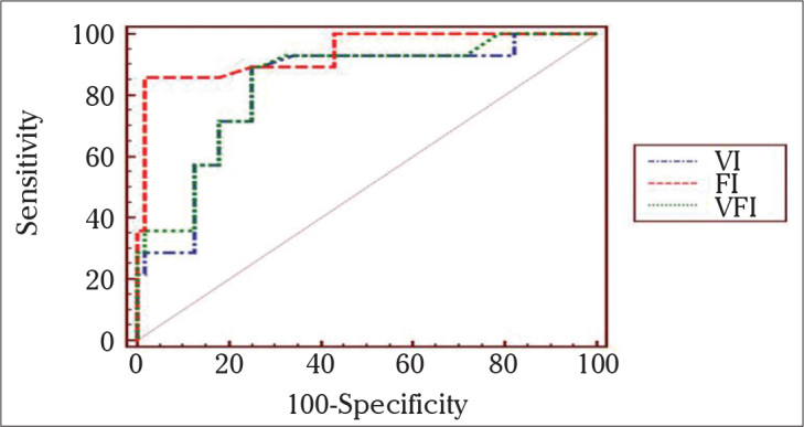
Results: Of the 84 women included in the study, 56 (66.7%) had benign endometrial lesions, and 28 (33.3%) had malignant endometrial lesions. Endometrial thickness, endometrial volume, and flow indices (VI, FI, and VFI) were higher in patients with malignant endometrium than those with benign endometrium. The area under the receiver operator characteristic curve (AUC) of endometrial thickness was 0.83, that of endometrial volume was 0.73, and that of the best power Doppler variable, FI, was 0.93. The best logistic regression model for predicting malignancy contained the variables endometrial thickness and FI; its AUC was 0.93.
Conclusion: The diagnostic performance of endometrial volume measured by 3D imaging with regard to discriminating between benign and malignant endometrium was not superior to that of endometrial thickness measured by 2D ultrasound examination, but 3D power Doppler flow indices are good diagnostic tools in predicting endometrial carcinoma.
Keywords: 3-dimensional ultrasound; Endometrial carcinoma; postmenopausal bleeding; power Doppler.
Figures
References
-
- Epstein E, Valentin L. Managing women with post-menopausal bleeding. Best Pract Res Clin Obstet Gynaecol. 2004;18:125–43. - PubMed
-
- Kanat-Pektas M, Gungor T, Mollamahmutoglu L. The evaluation of endometrial tumors by transvaginal and Doppler ultrasonography. Arch Gynecol Obstet. 2008;277:495–9. - PubMed
-
- Merce LT, Alcazar JL, Engels V, Troyano J, Bau S, Bajo JM. Endometrial volume and vascularity measurements by transvaginal three-dimensional ultrasonography and power Doppler angiography in stimulated and tumoral endometria: intraobserver reproducibility. Gynecol Oncol. 2006;100:544–50. - PubMed
-
- Makled AK, Elmekkawi SF, El-Refaie TA, El-Sherbiny MA. Three-dimensional power Doppler and endometrial volume as predictors of malignancy in patients with postmenopausal bleeding. J Obstet Gynaecol Res. 2013;39:1045–51. - PubMed
-
- Song HH. Analysis of correlated ROC areas in diagnostic testing. Biometrics. 1997;53:370–82. - PubMed
LinkOut - more resources
Full Text Sources
Other Literature Sources