Review
. 2014 Jun 28;6(6):284-300.
doi: 10.4329/wjr.v6.i6.284.

A handy review of carpal tunnel syndrome: From anatomy to diagnosis and treatment

Affiliations
Review

A handy review of carpal tunnel syndrome: From anatomy to diagnosis and treatment

Mohammad Ghasemi-Rad et al. World J Radiol. .

Abstract

Carpal tunnel syndrome (CTS) is the most commonly diagnosed disabling condition of the upper extremities. It is the most commonly known and prevalent type of peripheral entrapment neuropathy that accounts for about 90% of all entrapment neuropathies. This review aims to provide an outline of CTS by considering anatomy, pathophysiology, clinical manifestation, diagnostic modalities and management of this common condition, with an emphasis on the diagnostic imaging evaluation.

Keywords: Anatomy; Carpal tunnel syndrome; Computed tomography; Diagnosis; Magnetic resonance imaging; Nerve conduction study; Treatment; Ultrasonography.


Figures
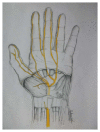
Figure 1
Figure 1
Sketch of the palm, showing specific details of the inner structures of the carpal tunnel (inside the wrist). The median nerve and its branches after the wrist are marked in yellow.
Figure 2
Figure 2
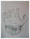
Sketch of the cross-section of the carpal tunnel on a hand. Median nerve is shown in yellow and the nine flexor tendons are marked in blue.
Figure 3
Figure 3
Axial computed tomography scan shows bony part of carpal tunnel at the level of outlet. Bony structures from left to right are HAMATE, CAPITATE, TRAPEZOID, TRAPEZIUM. FR (arrow) b and flexor tendons can be detected by computed tomography scan.
Figure 4
Figure 4
Axial ultrasound image shows flexor retinaculum bowing as an echogenic line (arrow) in carpal tunnel and cross sectional area of median nerve (stellate) in a patient with carpal tunnel syndrome.
Figure 5
Figure 5
Longitudinal color Doppler sonogram in a 40-year-old woman with severe carpal tunnel syndrome shows intraneural hypervascularity in the median nerve.
Figure 6
Figure 6
Spectral Doppler waveform of the median nerve shows low resistance hypervascularity of affected median nerve in a 40-year-old woman with severe carpal tunnel syndrome.
Figure 7
Figure 7
Axial ultrasound image shows hypoechoic cable like neural fascicle (arrows) separated by substratum hyperechoic fat in a patient with secondary carpal tunnel syndrome due to lipofibromatous hamartoma of the median nerve.
Figure 8
Figure 8
Axial T1W image of carpal tunnel at the level of tunnel outlet shows bony part of carpal tunnel as intermediate signal intensity composed from left to right hamate, capitates, trapezoid, trapezium. White arrow shows hook of hamate, yellow arrow shows median nerve, green arrow shows flexor retinaculum. Asterisks indicate carpal bones.

References

    1. Katz JN, Simmons BP. Clinical practice. Carpal tunnel syndrome. N Engl J Med. 2002;346:1807–1812. - PubMed
    1. Aroori S, Spence RA. Carpal tunnel syndrome. Ulster Med J. 2008;77:6–17. - PMC - PubMed
    1. Ibrahim I, Khan WS, Goddard N, Smitham P. Carpal tunnel syndrome: a review of the recent literature. Open Orthop J. 2012;6:69–76. - PMC - PubMed
    1. Mashoof AA, Levy HJ, Soifer TB, Miller-Soifer F, Bryk E, Vigorita V. Neural anatomy of the transverse carpal ligament. Clin Orthop Relat Res. 2001;(386):218–221. - PubMed
    1. Uchiyama S, Itsubo T, Nakamura K, Kato H, Yasutomi T, Momose T. Current concepts of carpal tunnel syndrome: pathophysiology, treatment, and evaluation. J Orthop Sci. 2010;15:1–13. - PubMed