The limbic degradation of aging brain: a quantitative analysis with diffusion tensor imaging
- PMID: 24977184
- PMCID: PMC4009154
- DOI: 10.1155/2014/196513
The limbic degradation of aging brain: a quantitative analysis with diffusion tensor imaging
Abstract
Introduction: The limbic system primarily responsible for our emotional life and memories is known to undergo degradation with aging and diffusion tensor imaging (DTI) is capable of revealing the white matter integrity. The aim of this study is to investigate age-related changes of quantitative diffusivity parameters and fiber characteristics on limbic system in healthy volunteers.
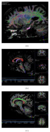
Methods: 31 healthy subjects aged 25-70 years were examined at 1,5 TMR. Quantitative fiber tracking was performed of fornix, cingulum, and the parahippocampal gyrus. The fractional anisotropy (FA) and apparent diffusion coefficient (ADC) measurements of bilateral hippocampus, amygdala, fornix, cingulum, and parahippocampal gyrus were obtained as related components.
Results: The FA values of left hippocampus, bilateral parahippocampal gyrus, and fornix showed negative correlations with aging. The ADC values of right amygdala and left cingulum interestingly showed negative relation and the left hippocampus represented positive relation with age. The cingulum showed no correlation. The significant relative changes per decade of age were found in the cingulum and parahippocampal gyrus FA measurements.
Conclusion: Our approach shows that aging affects hippocampus, parahippocampus, and fornix significantly but not cingulum. These findings reveal age-related changes of limbic system in normal population that may contribute to future DTI studies.
Figures


References
-
- Papez JW. A proposed mechanism of emotion. Archives of Neurology & Psychiatry. 1937;38(4):725–743.
-
- Kuzniecky R, Bilir E, Gilliam F, Faught E, Martin R, Hugg J. Quantitative MRI in temporal lobe epilepsy: evidence for fornix atrophy. Neurology. 1999;53(3):496–501. - PubMed
-
- Kodama F, Ogawa T, Sugihara S, et al. Transneuronal degeneration in patients with temporal lobe epilepsy: evaluation by MR imaging. European Radiology. 2003;13(9):2180–2185. - PubMed
-
- Bernasconi N, Duchesne S, Janke A, Lerch J, Collins DL, Bernasconi A. Whole-brain voxel-based statistical analysis of gray matter and white matter in temporal lobe epilepsy. NeuroImage. 2004;23(2):717–723. - PubMed
-
- Callen DJA, Black SE, Gao F, Caldwell CB, Szalai JP. Beyond the hippocampus: MRI volumetry confirms widespread limbic atrophy in AD. Neurology. 2001;57(9):1669–1674. - PubMed
MeSH terms
LinkOut - more resources
Full Text Sources
Other Literature Sources
Medical

