. 2014 Jul 30;5(14):5335-49.
doi: 10.18632/oncotarget.2104.

Galectin-4 expression is associated with reduced lymph node metastasis and modulation of Wnt/β-catenin signalling in pancreatic adenocarcinoma

Affiliations

Galectin-4 expression is associated with reduced lymph node metastasis and modulation of Wnt/β-catenin signalling in pancreatic adenocarcinoma

Mina Maftouh et al. Oncotarget. .

Abstract

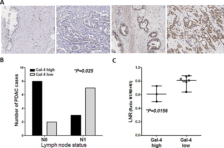
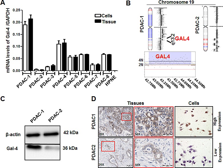
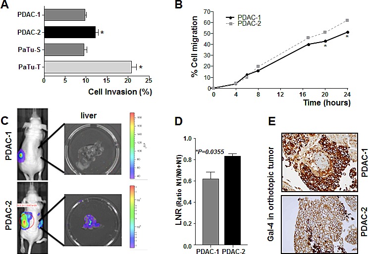
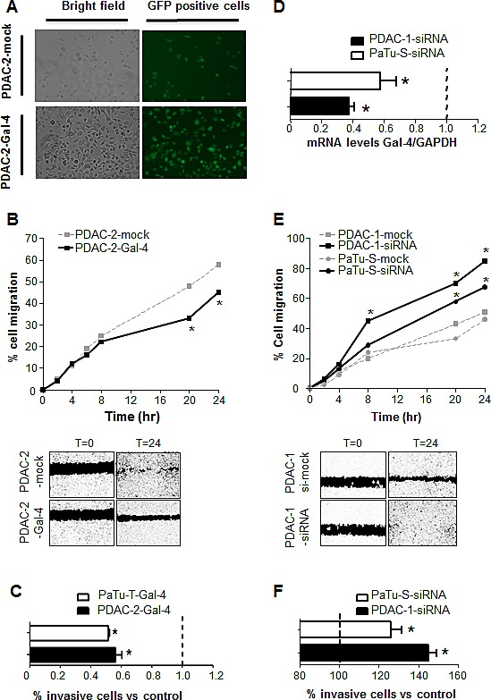
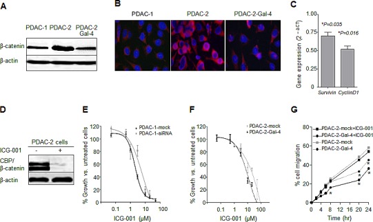
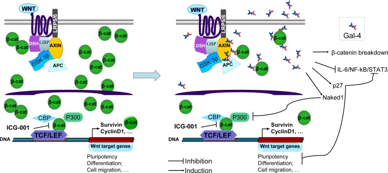
Galectin-4 (Gal-4) has been recently identified as a pivotal factor in the migratory capabilities of a set of defined pancreatic ductal adenocarcinoma (PDAC) cell lines using zebrafish as a model system. Here we evaluated the expression of Gal-4 in PDAC tissues selected according to their lymph node metastatic status (N0 vs. N1), and investigated the therapeutic potential of targeting the cross-link with the Wnt signaling pathway in primary PDAC cells. Analysis of Gal-4 expression in PDACs showed high expression of Gal-4 in 80% of patients without lymph node metastasis, whereas 70% of patients with lymph node metastases had low Gal-4 expression. Accordingly, in primary PDAC cells high Gal-4 expression was negatively associated with migratory and invasive ability in vitro and in vivo. Knockdown of Gal-4 in primary PDAC cells with high Gal-4 expression resulted in significant increase of invasion (40%) and migration (50%, P<0.05), whereas enforced expression of Gal-4 in primary cells with low Gal-4 expression reduced the migratory and invasive behavior compared to the control cells. Gal-4 markedly reduces β-catenin levels in the cell, counteracting the function of Wnt signaling, as was assessed by down-regulation of survivin and cyclin D1. Furthermore, Gal-4 sensitizes PDAC cells to the Wnt inhibitor ICG-001, which interferes with the interaction between CREB binding protein (CBP) and β-catenin. Collectively, our data suggest that Gal-4 lowers the levels of cytoplasmic β-catenin, which may lead to lowered availability of nuclear β-catenin, and consequently diminished levels of nuclear CBP-β-catenin complex and reduced activation of the Wnt target genes. Our findings provide novel insights into the role of Gal-4 in PDAC migration and invasion, and support the analysis of Gal-4 for rational targeting of Wnt/β-catenin signaling in the treatment of PDAC.


Figures

Figure 1
Figure 1. Patients with PDACs that highly express Gal-4 have a significantly decreased number of malignant cells in the lymph nodes, compared to patients with low Gal-4-PDACs
(A) Representative pictures of immunohistochemical analysis for Gal-4 expression in PDAC patients, showing differential Gal-4 expression (negative, weak, intermediate, strong). (B) Patients were classified in two groups, i.e. with (N1) or without (N0) lymph node metastasis. IHC analysis showed that eight patients without lymph node metastasis had high Gal-4 expression, while two patients had low Gal-4 expression, whereas in the group of patients with lymph node metastasis three patients had high Gal-4 expression while seven patients had low Gal-expression. (C) Analysis of the LNR ratio in the group with lymph node metastasis (N1).
Figure 2
Figure 2. Gal-4 is differentially expressed in primary PDAC cell cultures, as well as in their originator tissues
(A) Gal-4 mRNA levels in primary tumor cultures (white bars), and their originator tissues (black bars), as determined by qRT-PCR. Columns and bars represent the arithmetic means ± SEM of two independent experiments performed in triplicate. (B) aCGH analysis of copy number variations in the Gal-4 gene within the cytoband 19q13.2 of PDAC-1 and PDAC-2 cells shows a copy number gain (4N) in PDAC-1 cells. Left shifts and red color indicate the deleted segments, while right shifts and blue color indicate the gains/amplifications. The complete aCGH database is available at Gene Expression Omnibus (GEO) with accession number GSE44587. (C) Representative blots of Gal-4 protein expression in the PDAC-1 and PDAC-2 cells. As a loading control β-actin levels are indicated. (D) Representative pictures of Gal-4 protein expression in the PDAC-1 and PDAC-2 originator tissues and primary cells, with insets at higher magnification to allow evaluation of the intracellular pattern. Original magnification, 40X.
Figure 3
Figure 3. Gal-4 expression correlates with invasive and migratory capabilities of PDAC cells
(A) Invasion of primary PDAC-1 and PDAC-2 cells characterized by high and low Gal-4 expression, respectively, as measured by migration over collagen-coated transwell chambers. Columns and bars represent the means ± SEM of two independent experiments performed in triplicate. *P<0.05 (B) Migratory properties of PDAC-1 and PDAC-2 cells as determined in wound-healing assay. Points and bars represent the means ± SEM of two independent experiments performed in triplicate. *P<0.05 (C) Representative Firefly-luciferase bioluminescence images of orthotopic mouse models, derived from PDAC-1 and PDAC-2, characterized by low and high metastatic properties, respectively. (D) LNR ratio in PDAC-1 and PDAC-2 orthotopic mouse models. Columns and bars represent the means ± SEM values in three mice for each group. (E) Representative IHC pictures of Gal-4 protein expression in PDAC-1 and PDAC-2 orthotopic mouse models. Original magnification, 40X.
Figure 4
Figure 4. Modulation of Gal-4 expression alters the migratory and invasive behavior of PDAC cells
(A) Representative pictures of fluorescence microscopy in PDAC-2-Gal-4-GFP cells. (B) Migratory properties of PDAC-2-Gal-4 cells, as determined in wound-healing assay. Points represent the means of three independent experiments performed in triplicate. *P<0.05. The photograph under the graph shows a representative picture at 24 hours. (C) Invasion of PDAC-2-Gal-4 and PaTu-T-Gal-4 cells, as measured by migration over collagen-coated transwell chambers. Data are expressed as percentage of invading cells compared to mock transduced PDAC-2 and PaTu-T cells, respectively. Columns and bars represent the means ± SEM of three independent experiments performed in triplicate. *P<0.05. (D) mRNA levels of Gal-4 in PDAC-1 and PaTu-S, both transfected with Gal4-siRNA, as determined by qRT-PCR. Columns and bars represent the means ± SEM of three independent experiments performed in triplicate. *P<0.05. (E) Migratory properties of PDAC-1 and PaTu-S cells both transfected with Gal-4 siRNA. Points represent the means of three independent experiments performed in triplicate. *P<0.05. The photograph under the graph shows a representative picture at 24 hours. (F) Invasion of PDAC-1-siRNA and PaTu-S-siRNA cells, as measured by migration over collagen-coated transwell chambers. Data are expressed as percentage of invading cells compared to mock treated PDAC-1 and PaTu-S cells, respectively. Columns and bars represent the means ± SEM of three independent experiments performed in triplicate. *P<0.05.
Figure 5
Figure 5. Gal-4 modulates β-catenin levels and sensitizes PDAC cells to the Wnt inhibitor ICG-001
(A) Representative pictures of β-catenin protein expression as detected by Western blot in PDAC-1, PDAC-2 and PDAC-2-Gal-4 cells. As a loading control β-actin levels are indicated. (B) Immunofluorescence analysis of the accumulation of β-catenin (red) into the nucleus (blue), in PDAC-1, PDAC-2 and PDAC-2-Gal-4 cells. (C) mRNA levels of survivin and cyclin-D1 in PDAC-2-Gal-4 cells, as detected by quantitative RT-PCR. The results are calculated with the ΔCt method compared to mRNA levels of PDAC2 cells (set at 1). Columns and bars represent the means ± SEM of two independent experiments performed in triplicate. (D) β-catenin protein levels in PDAC-2 cells, untreated versus treated with ICG-001, as assessed by immunoprecipitation. (E) Inhibition of cell proliferation in PDAC-1-mock and PDAC-1-Gal4-siRNA after 72 hours exposure to the Wnt inhibitor ICG-001. Points and bars represent the means ± SEM of three independent experiments performed in triplicate. *P<0.05. (F) Inhibition of cell proliferation in PDAC-2-mock and PDAC-2-Gal-4 cells, after 72 hours exposure to the Wnt inhibitor ICG-001. Points and bars represent the means ± SEM of three independent experiments performed in triplicate. *P<0.05. (G) Migratory properties of PDAC-2-mock and PDAC-2-Gal-4 cells exposed to ICG-001 at IC50 concentration, as determined in wound-healing assay. Points represent the means of two independent experiments performed in triplicate. *P<0.05.
Figure 6
Figure 6. Model for Gal-4 effects on canonical Wnt signalling
Upon activation of the canonical Wnt signaling pathway, Frizzled (dark blue line) and low-density lipoprotein receptor-related protein 5/6 (LRP5/6) activate the protein Dishevelled (DSH), leading to Axin recruitment. The β-catenin (β-cat) “destruction complex”, composed of the proteins Axin, adenomatous polyposis coli (APC), glycogen synthase kinase-3 (GSK-3β) and GSK3-binding protein (GBP), is not able to phosphorylate β-cat, resulting in its accumulation and translocation into the nucleus. Interaction of nuclear β-cat with CREB-binding protein (CBP) leads to an active transcriptional complex for downstream target genes by binding to T-cell factor (TCF)- and lymphoid enhancer-binding protein (LEF)-family transcription factors. The Wnt inhibitor small molecule ICG-001 specifically binds to CBP thereby disrupting the interaction of CBP with β-cat. In the presence of Gal-4, overall β-cat levels in the cell are decreased and thus less ICG-001 is required to disrupt the interaction of CBP with β-catenin. Gal-4 can bind to axin, β-cat and APC, as shown in CRC [15]. Possibly, Gal-4 cross-links these components, thereby stabilizing the destruction complex, and enhancing degradation of β-cat. Furthermore, Gal-4 is shown to inhibit the IL-6/NF-kB/STAT3 pathway [11], to induce expression of the Wnt signaling inhibitor protein Naked1, and is involved in maintaining p27 levels in CRC [15, 43, 44]. Collectively, these effects due to involvement of Gal-4 might contribute to reduced cell migration.

Similar articles

Cited by

References

    1. Vincent A, Herman J, Schulick R, Hruban RH, Goggins M. Pancreatic cancer. Lancet. 2011;378(9791):607–620. - PMC - PubMed
    1. Hidalgo M. New insights into pancreatic cancer biology. Ann Oncol. 2012;23(Suppl 10):x135–138. - PubMed
    1. Valastyan S, Weinberg RA. Tumor metastasis: molecular insights and evolving paradigms. Cell. 2011;147(2):275–292. - PMC - PubMed
    1. Cummings RD, Liu FT. Varki A, Cummings RD, Esko JD, Freeze HH, Stanley P, Bertozzi CR, Hart GW, Etzler ME. Essentials of Glycobiology. 2. Cold Spring Harbor (NY): Cold Spring Harbor Laboratory Press; 2009. Galectins. Chapter 33. - PubMed
    1. Barondes SH, Castronovo V, Cooper DN, Cummings RD, Drickamer K, Feizi T, Gitt MA, Hirabayashi J, Hughes C, Kasai K, et al. Galectins: a family of animal beta-galactoside-binding lectins. Cell. 1994;76(4):597–598. - PubMed

Publication types