. 2014 Jun 27;6(3):1267-97.
doi: 10.3390/cancers6031267.

Merkel cell polyomavirus: molecular insights into the most recently discovered human tumour virus

Affiliations

Merkel cell polyomavirus: molecular insights into the most recently discovered human tumour virus

Gabrielė Stakaitytė et al. Cancers (Basel). .

Abstract

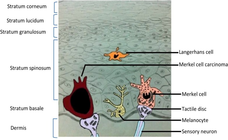
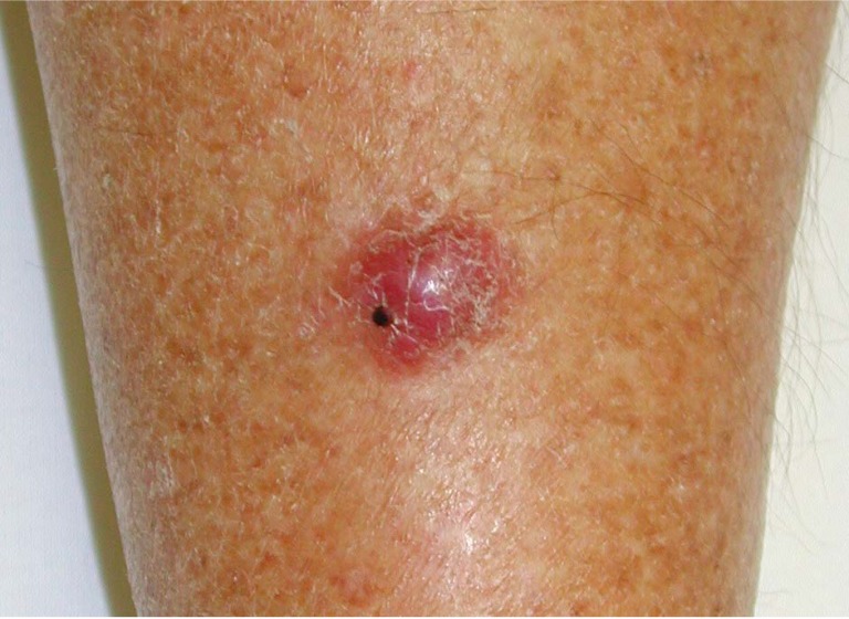
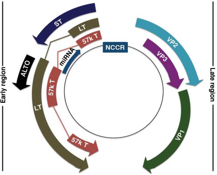
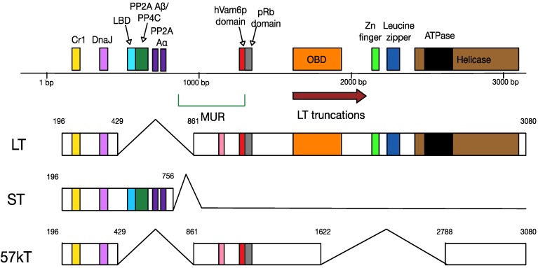
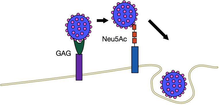
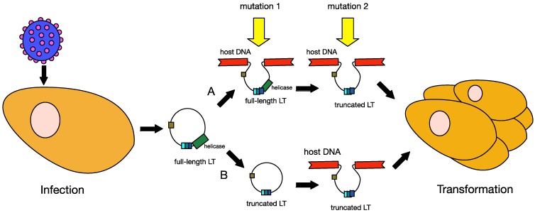
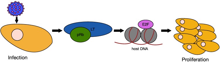
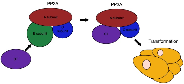
A fifth of worldwide cancer cases have an infectious origin, with viral infection being the foremost. One such cancer is Merkel cell carcinoma (MCC), a rare but aggressive skin malignancy. In 2008, Merkel cell polyomavirus (MCPyV) was discovered as the causative agent of MCC. It is found clonally integrated into the majority of MCC tumours, which require MCPyV oncoproteins to survive. Since its discovery, research has begun to reveal the molecular virology of MCPyV, as well as how it induces tumourigenesis. It is thought to be a common skin commensal, found at low levels in healthy individuals. Upon loss of immunosurveillance, MCPyV reactivates, and a heavy viral load is associated with MCC pathogenesis. Although MCPyV is in many ways similar to classical oncogenic polyomaviruses, such as SV40, subtle differences are beginning to emerge. These unique features highlight the singular position MCPyV has as the only human oncogenic polyomavirus, and open up new avenues for therapies against MCC.


Figures

Figure 1
Figure 1
Cross section of skin. Illustration of the six layers of skin, from the dermis to the stratum corneum. Examples of residents cells are included, among them a healthy Merkel cell and a cell that is part of a Merkel cell carcinoma tumour. Merkel cells are located in the stratum basale.
Figure 2
Figure 2
Merkel cell carcinoma. A typical MCC nodule present on the leg of a patient. Image credit to Howard Peach, Leeds Teaching Hospitals NHS Trust, UK.
Figure 3
Figure 3
MCPyV genome organisation. Non-coding control region (NCCR): bipartite origin of replication. Early gene region: Large T antigen (LT), small T antigen (ST), 57kT antigen (57kT), alternative T antigen open reading frame (ALTO), microRNA (miRNA). Late gene region: capsid proteins (VP1-3).
Figure 4
Figure 4
Mapping of the multiply-spliced MCPyV T antigens. The three T antigens are LT, ST and 57kT. All three encode CR1 (yellow, LXXLL) and DnaJ (lilac, HPDKGG) domains. ST also contains two PP2A Aα binding sites (R7 and L142), a PP2A Aβ/PP4C binding site (amino acids 97–111) and an large T-stabilisation domain (LSD, amino acids 91–95). LT shares the pRb binding domain with 57kT; in addition, it has unique origin binding (OBD), zinc finger, leucine zipper, ATPase and helicase domains. The MCPyV-unique region (MUR) of LT contains the hVam6p binding site.
Figure 5
Figure 5
Two-step attachmentandentry process of MCPyV. GAG—glycosaminoglycan, such as heparan sulfate. Neu5Ac—ganglioside with a linear Neu5Ac-α2,3-Gal motif.
Figure 6
Figure 6
NF-κB signaling. The IKK is activated after PAMP recognition by PRRs. This leads to the proteasomal degradation of IκB and release of NF-κB, which can then translocate into the nucleaus and activate the transcription of genes that have functions in the innate immunity.
Figure 7
Figure 7
Models of MCPyV-induced MCC tumourigenesis. MCPyV infection is thought to occur early in childhood of most people. Before tumourigenesis can occur, loss immunosurveillance must lead to proliferation of the virus. At least two mutations are needed before MCPyV can transform cells. In model A, the first mutation is thought to be the integration of the full-length viral genome into host DNA, while the second mutation is the truncation of LT. In model B, truncation of LT is thought to occur before integration. Either way, these changes in the virus lead to cellular transformation and tumour proliferation.
Figure 8
Figure 8
Effect of LT on cell proliferation. Upon MCPyV infection and T antigen expression, LT binds the regulatory protein pRb, thus inactivating it. This allows E2F to activate the transcription of cell cycle progression-associated genes, which switches the cell into S-phase and leads to cell proliferation.
Figure 9
Figure 9
Interaction of ST with PP2A. Polyomavirus ST competes with the B subunit of PP2A for binding to the structural A subunit and the catalytic C subunit. In SV40, this interaction between ST and PP2A Aα leads to cell transformation. Although the interaction of MCPyV ST with PP2A Aα may not be necessary for transformation, interaction with PP2A Aβ or the related PP4C may play a role.

Similar articles

Cited by

References

    1. Parkin D.M. The global health burden of infection-associated cancers in the year 2002. Int. J.Cancer. 2006;118:3030–3044. doi: 10.1002/ijc.21731. - DOI - PubMed
    1. Javier R.T., Butel J.S. The History of tumor virology. Cancer Res. 2008;68:7693–7706. doi: 10.1158/0008-5472.CAN-08-3301. - DOI - PMC - PubMed
    1. Moore P.S., Chang Y. Why do viruses cause cancer? Highlights of the first century of human tumour virology. Nat. Rev. Cancer. 2010;10:878–889. - PMC - PubMed
    1. Stewart S.E., Eddy B.E., Borgese N. Neoplasms in mice inoculated with a tumor agent carried in tissue culture. J. Natl. Cancer Inst. 1958;20:1223–1243. - PubMed
    1. Sweet B.H., Hilleman M.R. The Vacuolating Virus, SV40. Exp. Biol. Med. 1960;105:420–427. doi: 10.3181/00379727-105-26128. - DOI - PubMed

LinkOut - more resources