Review
. 2014 May;9(7):1063-82.
doi: 10.2217/nnm.14.55.

Targeted radiotherapy with gold nanoparticles: current status and future perspectives

Affiliations
Review

Targeted radiotherapy with gold nanoparticles: current status and future perspectives

Wilfred Ngwa et al. Nanomedicine (Lond). 2014 May.

Abstract

Radiation therapy (RT) is the treatment of cancer and other diseases with ionizing radiation. The ultimate goal of RT is to destroy all the disease cells while sparing healthy tissue. Towards this goal, RT has advanced significantly over the past few decades in part due to new technologies including: multileaf collimator-assisted modulation of radiation beams, improved computer-assisted inverse treatment planning, image guidance, robotics with more precision, better motion management strategies, stereotactic treatments and hypofractionation. With recent advances in nanotechnology, targeted RT with gold nanoparticles (GNPs) is actively being investigated as a means to further increase the RT therapeutic ratio. In this review, we summarize the current status of research and development towards the use of GNPs to enhance RT. We highlight the promising emerging modalities for targeted RT with GNPs and the corresponding preclinical evidence supporting such promise towards potential clinical translation. Future prospects and perspectives are discussed.

Keywords: cancer; gold nanoparticle; nanomedicine; radiotherapy; retinal disease.


Conflict of interest statement

Financial & competing interests disclosure

The authors have no other relevant affiliations or financial involvement with any organization or entity with a financial interest in or financial conflict with the subject matter or materials discussed in the manuscript apart from those disclosed.

No writing assistance was utilized in the production of this manuscript.

Figures

Figure 1
Figure 1. Third-generation gold nanoparticle platform for radiotherapy applications
(A) The synthetic strategy of the PEGylated gold nanoparticles followed by conjugation with AF647. (B) Transmission electron micrograph of the THPC-coated gold nanoparticles after PEGylation and (C) the confocal fluorescence imaging of HeLA cell cultures after uptake of fluorescent gold nanoparticles. AF647: Alexa Fluor 647; HV: High voltage; mag.: Magnification; mPEG: Monofunctional polyethylene gylcol; PEG: Polyethylene glycol; r.t.: Room temperature; THPC: Tetrakis(hydroxymethyl)phosphonium chloride. Reproduced with permission from [44] Translational Cancer Research.
Figure 2
Figure 2. In vitro DNA damage enhancement by gold nanoparticle during irradiation
(A) Surviving fraction of HeLA cells irradiated with 220 kVp x-rays (2 Gy) as a function of GNP concentration. (B) Comparison of unrepaired (residual) radiation damage for HeLA cells incubated with and without GNPs. The rDEF represents the ratio of the residual DNA damage with and without GNPs. The rDEF is shown for four sets of irradiation experiments (2.1–4.5 cGy/h), as well as for no irradiation (0 cGy/h). The figure is from Ngwa et al. [4]. (C) Evidence of dose enhancement during 6 MV external RT; damage enhancement as a function of depth in tissue-equivalent material for conventional (open-field) photon-field delivery with square apertures of sizes 5, 10 and 15 cm. (D) Damage enhancement as a function of depth in tissue-equivalent material for conventional (15 × 15 cm) and FFF (15 × 15 cm) photon-field delivery. FFF: Flattening filter free; PGNP: PEGylated gold nanoparticle; rDEF: Residual DNA damage enhancement factor. (A) Reproduced with permission from [44] Translational Cancer Research. (B) Reproduced with permission from [4]. (C & D) Reproduced with permission from [3].
Figure 3
Figure 3. Radiotherapy application with in situ dose painting
(A) 125I radiation-only dose distribution. (B) Dose distribution using the radiotherapy application with in situ dose painting approach where a gold-loaded RTB is used instead of usual inert spacer. (C) Not to scale. Tumor cell in high-risk ROI receives a dose boost due to 125I-induced emission of photo-/Auger electrons. (D) GNP release kinetics can be customized for sustained in situ radiosensitization (blue curve) compared with the current approach of weekly systemic administration of radiosensitizer (multiple peaks). GNP: Gold nanoparticle; ROI: Region of interest; RTB: Radiotherapy biomaterial.
Figure 4
Figure 4. Electron microscopy image (1 × 1 µM) of smart radiotherapy biomaterial loaded with stealth nanoparticles
The arrow shows a gold nanoparticle embedded in the polymer matrix indicated by the ‘X’.
Figure 5
Figure 5. Gold nanoparticle-aided Oraya Therapy™ (Oraya Therapeutics Inc., CA, USA) dose distribution
(A) Spatial map of Oraya Therapy™ (Oraya Therapeutics Inc., CA, USA) dose distribution (in Gy) within the eye of a patient with an anterior–posterior diameter of the eye of approximately 24 mm. (B) Idealistic spatial map of Oraya Therapy dose distribution illustrating a possible treatment scenario when using gold nanoparticles. In this scenario, the directly administered Oraya Therapy dose is reduced by a factor of greater than 3 (corresponding to dose enhancement factor level), while the dose to the choroidal neovasculature is maintained due to dose enhancement by targeted gold nanoparticles. Such a scenario would lead to significantly reduced dose to neighboring healthy tissue. Reproduced with permission from [101] © IOP Publishing.
Figure 6
Figure 6. Three of the emerging approaches of gold nanoparticle-aided radiotherapy
(A) Customizable radiotherapy enhancement with GNPs, (B) GNPs as vascular-distrupting agents and (C) radiotherapy application with in situ dose painting with GNPs. AMD: Age-related macular degeneration; GNP: Gold nanoparticle.

References

    1. Detappe A, Tsiamas P, Ngwa W, Zygmanski P, Makrigiorgos M, Berbeco R. The effect of flattening filter free delivery on endothelial dose enhancement with gold nanoparticles. Med. Phys. 2013;40(3):031706. - PMC - PubMed
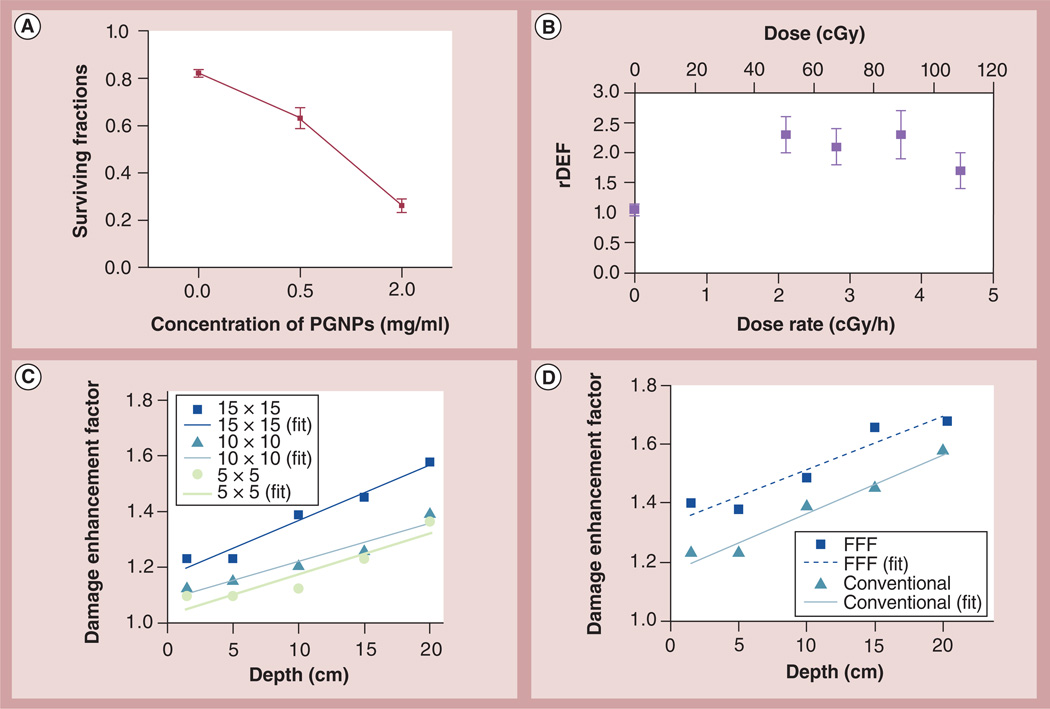
    1. Tsiamas P, Liu B, Cifter F, et al. Impact of beam quality on megavoltage radiotherapy treatment techniques utilizing gold nanoparticles for dose enhancement. Phys. Med. Biol. 2013;58(3):451–464. - PubMed
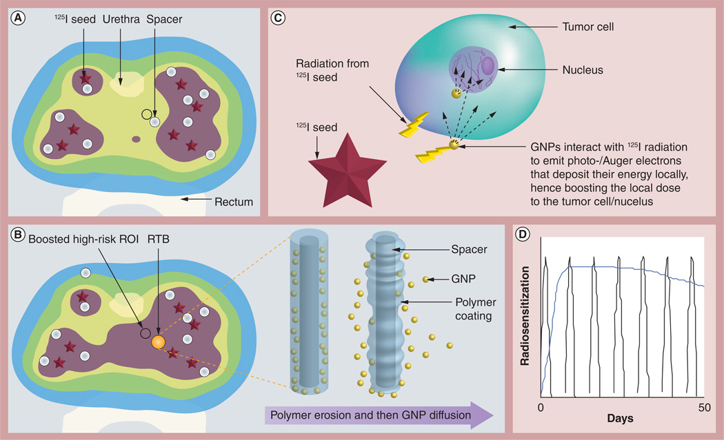
    1. Berbeco RI, Korideck H, Ngwa W, et al. DNA damage enhancement from gold nanoparticles for clinical MV photon beams. Radiat. Res. 2012;178(6):604–608. •• Provides experimental evidence of gold nanoparticle enhancement for different clinical MV photon beams. It is also the first experimental evidence of radiosensitization by gold nanoparticles during continuous low-dose-rate I125 brachytherapy.

    1. Ngwa W, Korideck H, Kassis AI, et al. In vitro radiosensitization by gold nanoparticles during continuous low-dose-rate gamma irradiation with I-125 brachytherapy seeds. Nanomedicine. 2013;9(1):25–27. - PMC - PubMed
    1. Chang JY, Zhang X, Wang X, et al. Significant reduction of normal tissue dose by proton radiotherapy compared with three-dimensional conformal or intensity-modulated radiation therapy in stage I or stage III non-small-cell lung cancer. Int. J. Radiat. Oncol. Biol. Phys. 2006;65(4):1087–1096. - PubMed

Publication types