

MRI-guided selection of patients for treatment of acute ischemic stroke
- PMID: 24978637
- PMCID: PMC4154317
- DOI: 10.1097/WCO.0000000000000110
MRI-guided selection of patients for treatment of acute ischemic stroke
Abstract
Purpose of review: To summarize what is known about the use of MRI in acute stroke treatment (predominantly thrombolysis), to examine the assumptions and theories behind the interpretation of magnetic resonance images of acute ischemic stroke and how they are used to select patients for therapies, and to suggest directions for future research.
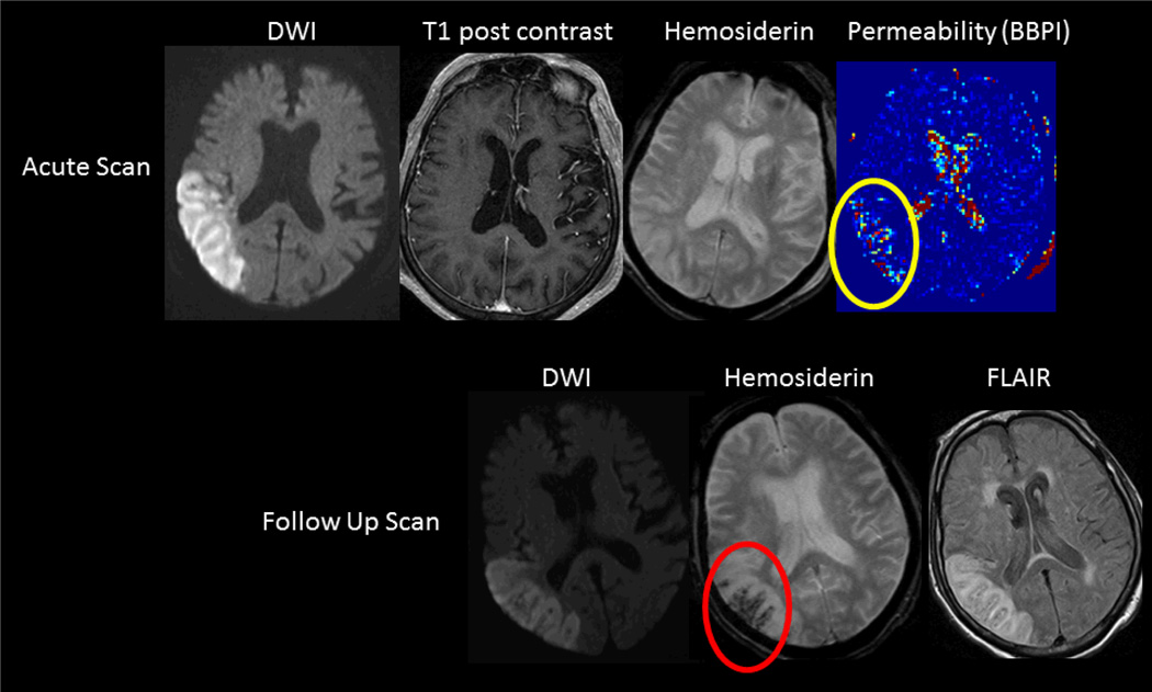
Recent findings: Recent studies have been contradictory about the usefulness of MRI in selecting patients for treatment. New MRI models for selecting patients have emerged that focus not only on the ischemic penumbra but also on the infarct core. Fixed time-window selection parameters are being replaced by timing-based individualized MRI stroke features. New ways to interpret traditional MRI stroke sequences are emerging.
Summary: Although the efficacy of acute stroke treatment is time dependent, the use of fixed time windows cannot account for individual differences in infarct evolution, which could potentially be detected with MRI. Although MRI shows promise for identifying patients who should be treated, as well as excluding patients who should not be treated, definitive evidence is still lacking. Future research should focus on validating the use of MRI to select patients for intravenous therapies in extended time windows.
Conflict of interest statement
Conflicts-of-interest/Disclosures:
There are no disclosures or conflicts-of-interest.
Figures


References
-
- Adams HP, Jr, del Zoppo G, Alberts MJ, Bhatt DL, Brass L, Furlan A, et al. Guidelines for the early management of adults with ischemic stroke: a guideline from the American Heart Association/American Stroke Association Stroke Council, Clinical Cardiology Council, Cardiovascular Radiology and Intervention Council, and the Atherosclerotic Peripheral Vascular Disease and Quality of Care Outcomes in Research Interdisciplinary Working Groups: the American Academy of Neurology affirms the value of this guideline as an educational tool for neurologists. Stroke. 2007;38(5):1655–1711. - PubMed
-
- Astrup J, Siesjo BK, Symon L. Thresholds in cerebral ischemia - the ischemic penumbra. Stroke. 1981;12(6):723–725. - PubMed
-
- Schlaug G, Benfield A, Baird AE, Siewert B, Lovblad KO, Parker RA, et al. The ischemic penumbra: operationally defined by diffusion and perfusion MRI. Neurology. 1999;53(7):1528–1537. - PubMed
-
- Tissue plasminogen activator for acute ischemic stroke. The National Institute of Neurological Disorders and Stroke rt-PA Stroke Study Group. NEnglJMed. 1995;333(24):1581–1587. - PubMed
Publication types
MeSH terms
Grants and funding
LinkOut - more resources
Full Text Sources
Other Literature Sources
Medical
Research Materials
