. 2014 Jun 30;3(6):e107.
doi: 10.1038/oncsis.2014.19.

MUC1 regulates cyclin D1 gene expression through p120 catenin and β-catenin

Affiliations

MUC1 regulates cyclin D1 gene expression through p120 catenin and β-catenin

X Liu et al. Oncogenesis. .

Abstract

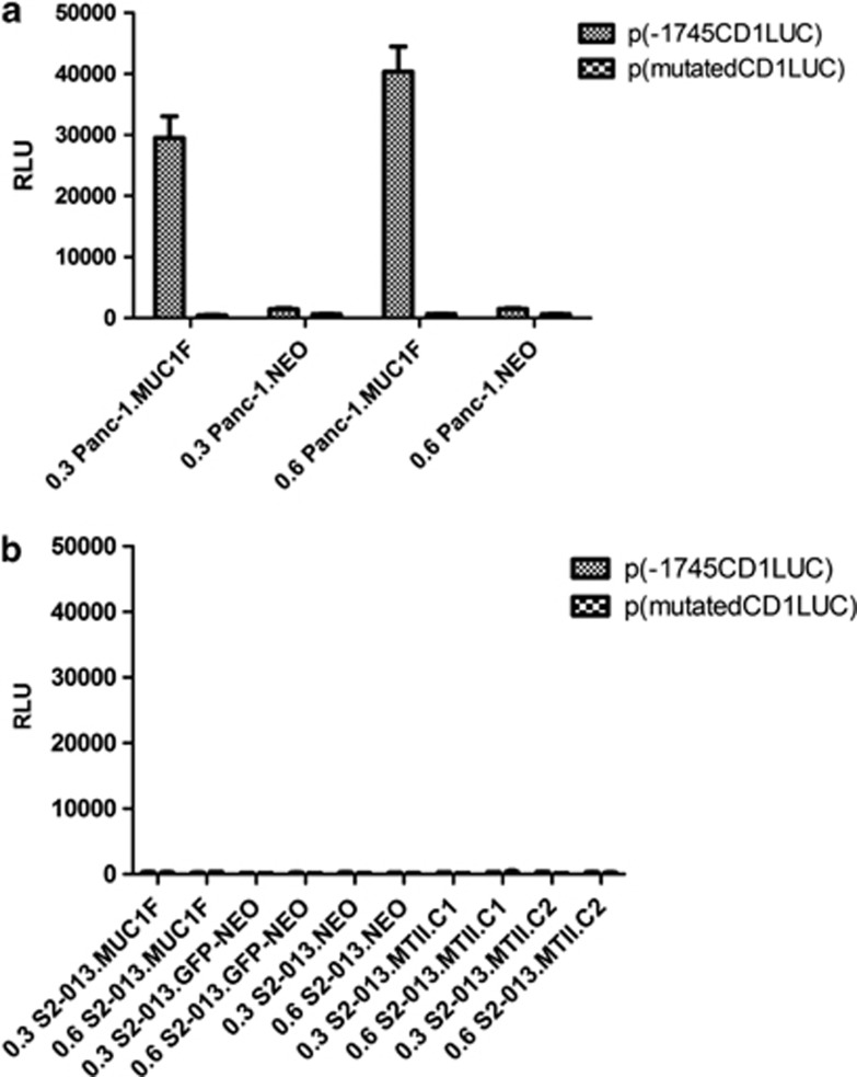
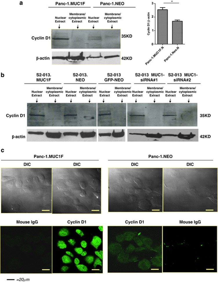
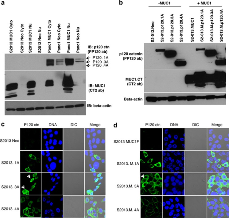
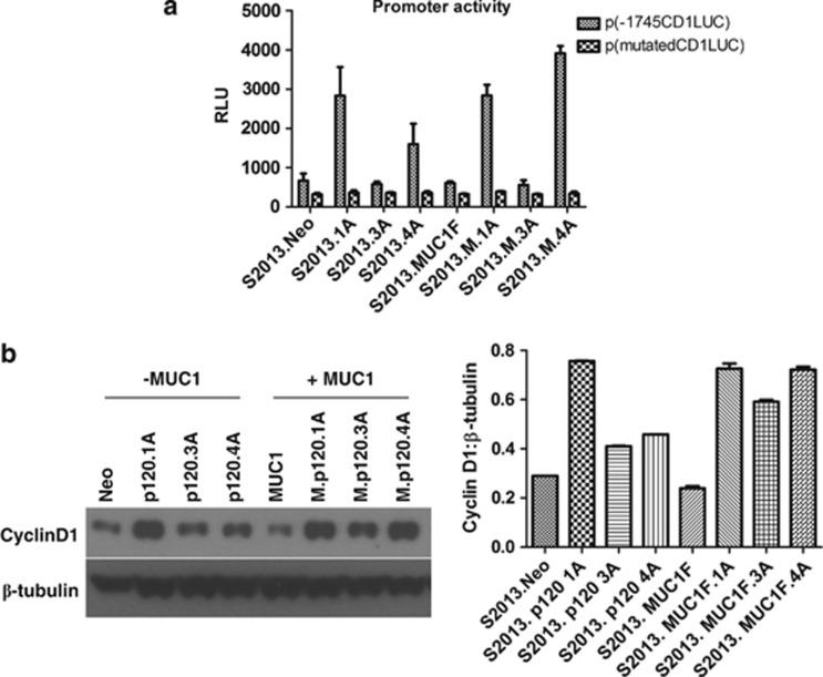
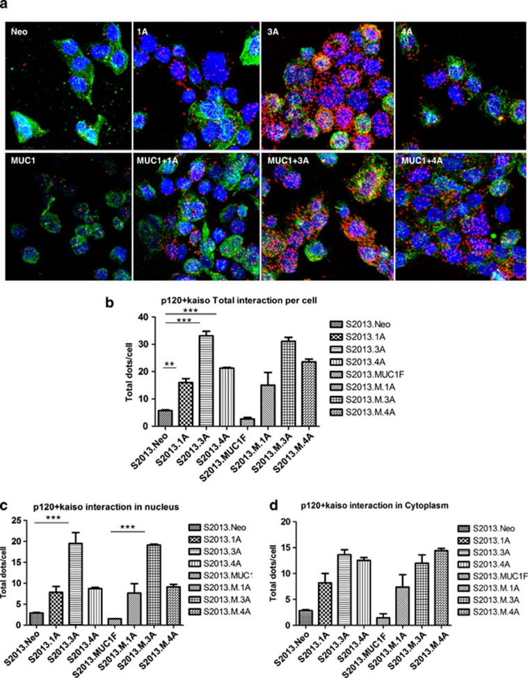
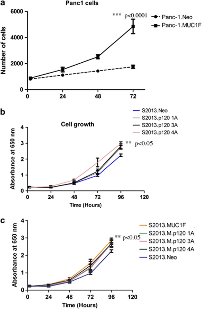
MUC1 interacts with β-catenin and p120 catenin to modulate WNT signaling. We investigated the effect of overexpressing MUC1 on the regulation of cyclin D1, a downstream target for the WNT/β-catenin signaling pathway, in two human pancreatic cancer cell lines, Panc-1 and S2-013. We observed a significant enhancement in the activation of cyclin D1 promoter-reporter activity in poorly differentiated Panc1.MUC1F cells that overexpress recombinant MUC1 relative to Panc-1.NEO cells, which express very low levels of endogenous MUC1. In stark contrast, cyclin D1 promoter activity was not affected in moderately differentiated S2-013.MUC1F cells that overexpressed recombinant MUC1 relative to S2-013.NEO cells that expressed low levels of endogenous MUC1. The S2-013 cell line was recently shown to be deficient in p120 catenin. MUC1 is known to interact with P120 catenin. We show here that re-expression of different isoforms of p120 catenin restored cyclin D1 promoter activity. Further, MUC1 affected subcellular localization of p120 catenin in association with one of the main effectors of P120 catenin, the transcriptional repressor Kaiso, supporting the hypothesis that p120 catenin relieved transcriptional repression by Kaiso. Thus, full activation of cyclin D1 promoter activity requires β-catenin activation of TCF-lef and stabilization of specific p120 catenin isoforms to relieve the repression of KAISO. Our data show MUC1 enhances the activities of both β-catenin and p120 catenin.


Figures

Figure 1
Figure 1
Luciferase assay detecting effect of MUC1 on cyclin D1 promoter activity. Cells were transfected with 300 or 600 ng of the reporter plasmids containing 1745 bp of the cyclin D1 gene promoter (−1745CD1Luc, TOPFLASH) consists of wild-type LEF-1/Tcf-4-binding site or reporter plasmids containing mutated LEF-1/Tcf-4-binding site in the cyclin D1 gene promoter (FOPFLASH), together with 400 ng of a synthetic Renilla luciferase reporter plasmids SV40 as transfection control. Each transfection was carried out in duplicate plates, and the data (relative lumenescence units-RLU) shown here are representative of three independent experiments. (a) Panc-1.MUC1F or Panc-1.Neo (95% confluence). (b) S2-013.MUC1F cells, S2-013.NEO cells, S2-013 GFP-NEO, two clones of S2-013 cells in which MUC1 was knocked down by MUC1-siRNA—S2-013.MTII.C1 and S2-013.MTII.C2 (95% confluence).
Figure 2
Figure 2
Western blot protein levels of cyclin D1 in pancreatic cancer cell lines. (a, b) Membrane/cytoplasmic extracts and nuclear extracts from Panc-1.MUC1F, Panc-1.NEO, S2-013.MUC1F, S2-013.NEO, S2-013 GFP-NEO, two clones of MUC1-siRNA S2-013 cells were subjected to 4–20% SDS–PAGE and analyzed by immunoblot (IB) with an anti-cyclin D1 mAb and β-actin as a loading control. Right panel in a shows densitometry analysis of the signal for nuclear cyclin D1 normalized to the signal for β-actin. *P<0.01, significant difference. (c) Cyclin D1 protein expression is enhanced by expression of MUC1 in Panc-1.MUC1F cells as compared with Panc-1.NEO cells. Confocal microscopy was used to determine the cyclin D1 protein expression in Panc-1.MUC1F and Panc-1.NEO cells. Cells grown above 90% confluence on coverslips were fixed with 4% paraformaldehyde and permeablized with Trixton X-100 before incubation with mAb anti-cyclin D1, which was identified with fluorescein isothiocyanate-conjugated secondary antibody and visualized as green color. Images were examined with a Zeiss LSM 410 laser scanning microscope (Bar=20 μM; × 100 magnification; results shown here represent three or four individual cell scanning observations.).
Figure 3
Figure 3
P120 catenin was not detected in S2-013 cells and re-expression of different p120 catenin isoforms in this cells showed distinct subcellular localization. (a) Membrane/cytoplasmic extracts and nuclear extracts from S2-013.NEO, S2-013.MUC1F, Panc-1.NEO, Panc-1.MUC1F were subjected to 10% SDS–PAGE and analyzed by immunoblot (IB) with an anti-p120 catenin mAb. The same blot was striped and reprobed with anti-MUC1.CT antibody. (b) Re-expression of different p120 catenin isoforms were confirmed by western blot using the same p120 catenin antibody and MUC1 antibody. (c and d) Immunofluorescence analysis of re-expression of p120 catenin in S2-013 cells. The green color indicates stains for p120 catenin. Blue indicates 4′-6-diamidino-2-phenylindole (DAPI) staining for nuclei. The arrowhead indicates subcellular localization of p120 catenin in the nucleus.
Figure 4
Figure 4
Re-expression of specific p120 catenin isoform restores cyclin D1 promoter activity and increases its protein expression level. (a) Luciferase reporter assay of cyclin D1 promoter. S2-013 cells with re-expression of different p120 catenin isoforms (with/without MUC1 expression) were transfected with wild-type cyclin D1 reporter construct, which contains wild-type LEF-1/Tcf-4-binding site or reporter plasmids containing mutated LEF-1/Tcf-4-binding site in the cyclin D1 gene promoter (FOPFLASH), together with Renilla luciferase reporter plasmids SV40 as transfection control. Each transfection was carried out in duplicate plates, and the data shown here are relative luminescence units (RLUs) derived from luminescence assays on cell extracts that are representative of three independent experiments. (b) Nuclear extracts from S2-013 cells with re-expression of different p120 catenin isoforms with or without MUC1 expression were subjected to 10% SDS–PAGE and analyzed by immunoblot (IB) with an anti-cyclin D1 catenin mAb or β-tubulin as a loading control. Right panel shows densitometry analysis of the signal for cyclin D1 normalized to the signal for β-tubulin.
Figure 5
Figure 5
Different p120 catenin isoforms associate with Kaiso. (a) Proximity ligation assay (PLA) was used to detect interactions between p120 catenin and Kaiso in S2-013 cells with re-expression of different p120 catenin isoforms with or without MUC1. The red dots indicate interactions between p120 catenin and Kaiso. Blue (4′-6-diamidino-2-phenylindole, DAPI) staining indicates nuclei. The green indicates α-tubulin. (bd) Quantification of results from PLAs. Results were compiled from three independent experiments.
Figure 6
Figure 6
Effect of MUC1 expression and p120 catenin re-expression on in vitro cell growth in Panc-1 and S2-013 cells. (a) In vitro growth rate of Panc-1.MUC1F and Panc-1.NEO cells were evaluated by counting cells as described in the Materials and methods. (b and c) Cell growth assay using a methylene blue cell dye to measure the in vitro growth rate of S2-013 cells with re-expression of different p120 catenin isoforms with or without MUC1 expression.

References

    1. McDermott KM, Crocker PR, Harris A, Burdick MD, Hinoda Y, Hayashi T, et al. Overexpression of MUC1 reconfigures the binding properties of tumor cells. Int J Cancer. 2001;94:783–791. - PubMed
    1. Rahn JJ, Chow JW, Horne GJ, Mah BK, Emerman JT, Hoffman P, et al. MUC1 mediates transendothelial migration in vitro by ligating endothelial cell ICAM-1. Clin Exp Metastasis. 2005;22:475–483. - PubMed
    1. Spicer AP, Duhig T, Chilton BS, Gendler SJ. Analysis of mammalian MUC1 genes reveals potential functionally important domains. Mamm Genome. 1995;6:885–888. - PubMed
    1. Singh PK, Behrens ME, Eggers JP, Cerny RL, Bailey JM, Shanmugam K, et al. Phosphorylation of MUC1 by Met modulates interaction with p53 and MMP1 expression. J Biol Chem. 2008;283:26985–26995. - PMC - PubMed
    1. Behrens ME, Grandgenett PM, Bailey JM, Singh PK, Yi CH, Yu F, et al. The reactive tumor microenvironment: MUC1 signaling directly reprograms transcription of CTGF. Oncogene. 2010;29:5667–5677. - PMC - PubMed