. 2014 Jul 30;5(14):5295-303.
doi: 10.18632/oncotarget.2092.

Loss of ARID1A expression sensitizes cancer cells to PI3K- and AKT-inhibition

Affiliations

Loss of ARID1A expression sensitizes cancer cells to PI3K- and AKT-inhibition

Eleftherios P Samartzis et al. Oncotarget. .

Abstract

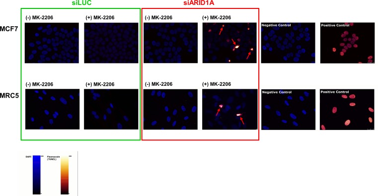
ARID1A mutations are observed in various tumors, including ovarian clear cell (OCCC) and endometrioid carcinomas, endometrial, and breast carcinomas. They commonly result in loss of ARID1A-protein expression and frequently co-occur with PI3K/AKT-pathway activating mechanisms. The aim of this study was to test the hypothesis as to whether PI3K/AKT-pathway activation is a critical mechanism in ARID1A-mutated tumors and if consequently ARID1A-deficient tumors show increased sensitivity to treatment with PI3K- and AKT-inhibitors. Upon ARID1A knockdown, MCF7 breast cancer cells and primary MRC5 cells exhibited a significantly increased sensitivity towards the AKT-inhibitors MK-2206 and perifosine, as well as the PI3K-inhibitor buparlisib. Knockdown of ARID1A in MCF7 led to an increase of pAKT-Ser473. AKT-inhibition with MK-2206 led to increased apoptosis and to a decrease of pS6K in ARID1A-depleted MCF7 cells but not in the controls. In five OCCC cell lines ARID1A-deficiency correlated with increased pAKT-Ser473 levels and with sensitivity towards treatment with the AKT-inhibitor MK-2206. In conclusion, ARID1A-deficient cancer cells demonstrate an increased sensitivity to treatment with small molecule inhibitors of the PI3K/AKT-pathway. These findings suggest a specific requirement of the PI3K/AKT pathway in ARID1A-deficient tumors and reveal a synthetic lethal interaction between loss of ARID1A expression and inhibition of the PI3K/AKT pathway.


Figures

Figure 1
Figure 1. Loss of ARID1A expression leads to increased sensitivity towards the AKT-inhibitors MK-2206 and perifosine as well as towards the PI3K-inhibitor buparlisib in MCF7 and MRC5 cells
(A) Increased sensitivity of ARID1A-depleted MCF7 and MRC5 cells towards the AKT-inhibitor MK-2206. (B) Increased sensitivity of ARID1A-depleted MCF7 and MRC5 cells towards the AKT-inhibitor perifosine. (C) Increased sensitivity of ARID1A-depleted MCF7 and MRC5 cells towards the PI3K-inhibitor buparlisib. The ARID1A-deficient OVSAYO cell line served as a control in all the experiments. The p-values indicate the divergence of the IC50-values calculated by an F-test. Significance was assumed for p<0.05 and is indicated in the figures (ns: not significant).
Figure 2
Figure 2. Loss of ARID1A expression increases vulnerability of cancer and primary cells to AKT-inhibition resulting in increased apoptosis
(A) Immunoblot showing an increased phosphorylation of AKT at Ser-473 in response to knockdown of ARID1A by siRNAs in the MCF7 cell line. ARID1A depletion increased pS6K downstream. Treatment with the AKT-inhibitor MK-2206 (at a concentration of 10−6M) completely abrogated pAKT-Ser473 in ARID1A-deficient MCF7 cells and led to reduced pS6K, in contrast to the controls where pS6K was not reduced. PARP-1 cleavage was markedly increased in ARID1A-deficient MCF7 cells treated with MK-2206 indicating an increased apoptosis rate, in contrast to the controls where no increase of the apoptosis rate was detectable after treatment with MK-2206. (B) Immunoblot demonstrating ARID1A knockdown in MRC5 cell line. The relative level of pAKT-Ser473 compared to the respective AKT level was increased in ARID1A-depleted MRC5 cells and completely abrogated by the treatment with the AKT-inhibitor MK-2206 (10−6M). (C) Immunoblot demonstrating the effects of a treatment with the AKT-inhibitor MK-2206 (10−6M) in the ARID1A-deficient OCCC cell line OVSAYO, which was used as a negative control for the knockdown experiments. Knockdown of ARID1A by siRNAs did not show an effect on pAKT-Ser473 and PARP-1 cleavage in this cell line, confirming that the effects are specifically due to the knockdown of the ARID1A gene. Combination of ARID1A-knockdown and treatment with MK-2206 did not cause increased PARP-1 cleavage in this control cell line. (D) MCF7 cells were seeded in 96-well plates and transfected with siLUC, siAKT1, siARID1A, or double-transfected with siAKT1 and siARID1A. Viable cells were counted with an MTS assay after 5 days (indicated as percent of viable cells in relation to the siLUC-transfected controls). ARID1A knockdown led to an increased proliferation of MCF7 cells in comparison to the controls. Knockdown of only AKT1 did reduce measurable pAKT-Ser473- and AKT- levels and led to a decreased level of pS6K (as shown in (E)), but did not lead to a difference in the amount of viable MCF7 cells. Combined knockdown with AKT1 in contrast completely abrogated the increased proliferation in ARID1A-depleted MCF7 cells. (E) Western blot showing the decreased expression of pAKT-Ser473, AKT, and pS6K 120h after AKT1-siRNA knockdown in MCF7 cells.
Figure 3
Figure 3. Treatment with the AKT-inhibitor MK-2206 causes apoptosis in ARID1A-depleted MCF7 and MRC5 cells
Treatment with MK-2206 (10−6M) for 48h led to an increased rate of apoptotic cells in ARID1A-depleted MCF7 and MRC5 cells compared to the controls. Note the different staining pattern of apoptotic cells compared to the positive controls, which is caused by pyknosis and/or fragmentation of the nuclei in the apoptotic cells (indicated by red arrows). The more homogeneous staining in the positive controls is the result of treatment of intact nuclei of fixated non-apoptotic cells with DNase immediately prior to the addition of the TUNEL incubation mix. Negative controls were obtained by omission of the TUNEL enzyme in the incubation mix. The nuclei are represented in blue (DAPI staining) and the fluorescein signal is labelled in red-glow.
Figure 4
Figure 4. Loss of ARID1A expression is associated with high sensitivity to the AKT-inhibitor MK-2206 in ovarian clear cell carcinoma cell lines
(A) The three ARID1A-deficient OCCC cell lines OVSAYO, OVISE, and HCH-1 were highly sensitive to a treatment with MK-2206 whereas the two OCCC cell lines ES-2 and RMG-1 with intact ARID1A expression were resistant to the same treatment. The logIC50 values significantly differed between ARID1A-intact and ARID1A-deficient OCCC cell lines (***p<0.0001) as verified with an F-test (Graph Pad Prism, version 6). (B) Immunoblot showing intact ARID1A expression in ES-2 and RMG-1 and loss of ARID1A expression in OVSAYO, OVISE, and HCH-1. Treatment with MK-2206 (10−6M) completely abrogated detectable pAKT-Ser473 levels in all OCCC cell lines. (C) Densitometric quantification of the relative pAKT-Ser473/AKT expression in the five untreated and treated OCCC cell lines (ImageJ version 1.46, NIH, USA). Relative pAKT-Ser473/AKT levels were significantly increased in the three ARID1A-deficient OCCC cell lines OVSAYO, OVISE, and HCH-1, as compared to the two ARID1A-intact OCCC cell lines ES-2 and RMG-1 (*p<0.01) as verified by a t-test (Graph Pad Prism, version 6).

References

    1. Shain AH, Pollack JR. The Spectrum of SWI/SNF Mutations, Ubiquitous in Human Cancers. PLoS One. 2013;8(1):e55119. - PMC - PubMed
    1. Wilson BG, Roberts CW. SWI/SNF nucleosome remodellers and cancer. Nat Rev Cancer. 2011;11(7):481–492. - PubMed
    1. Wiegand KC, Shah SP, Al-Agha OM, Zhao Y, Tse K, Zeng T, Senz J, McConechy MK, Anglesio MS, Kalloger SE, Yang W, Heravi-Moussavi A, Giuliany R, Chow C, Fee J, Zayed A, et al. ARID1A mutations in endometriosis-associated ovarian carcinomas. N Engl J Med. 2010;363(16):1532–1543. - PMC - PubMed
    1. Jones S, Wang TL, Shih IM, Mao TL, Nakayama K, Roden R, Glas R, Slamon D, Diaz LA, Vogelstein B, Kinzler KW, Velculescu VE, Papadopoulos N. Frequent mutations of chromatin remodeling gene ARID1A in ovarian clear cell carcinoma. Science. 2010;330(6001):228–231. - PMC - PubMed
    1. Wiegand KC, Lee AF, Al-Agha OM, Chow C, Kalloger SE, Scott DW, Steidl C, Wiseman SM, Gascoyne RD, Gilks B, Huntsman DG. Loss of BAF250a (ARID1A) is frequent in high-grade endometrial carcinomas. J Pathol. 2011;224(3):328–333. - PubMed

Publication types

MeSH terms