. 2014 Aug;184(8):2333-41.
doi: 10.1016/j.ajpath.2014.04.009. Epub 2014 Jun 27.

Association between periodontal disease and inflammatory arthritis reveals modulatory functions by melanocortin receptor type 3

Affiliations

Association between periodontal disease and inflammatory arthritis reveals modulatory functions by melanocortin receptor type 3

Trinidad Montero-Melendez et al. Am J Pathol. 2014 Aug.

Abstract

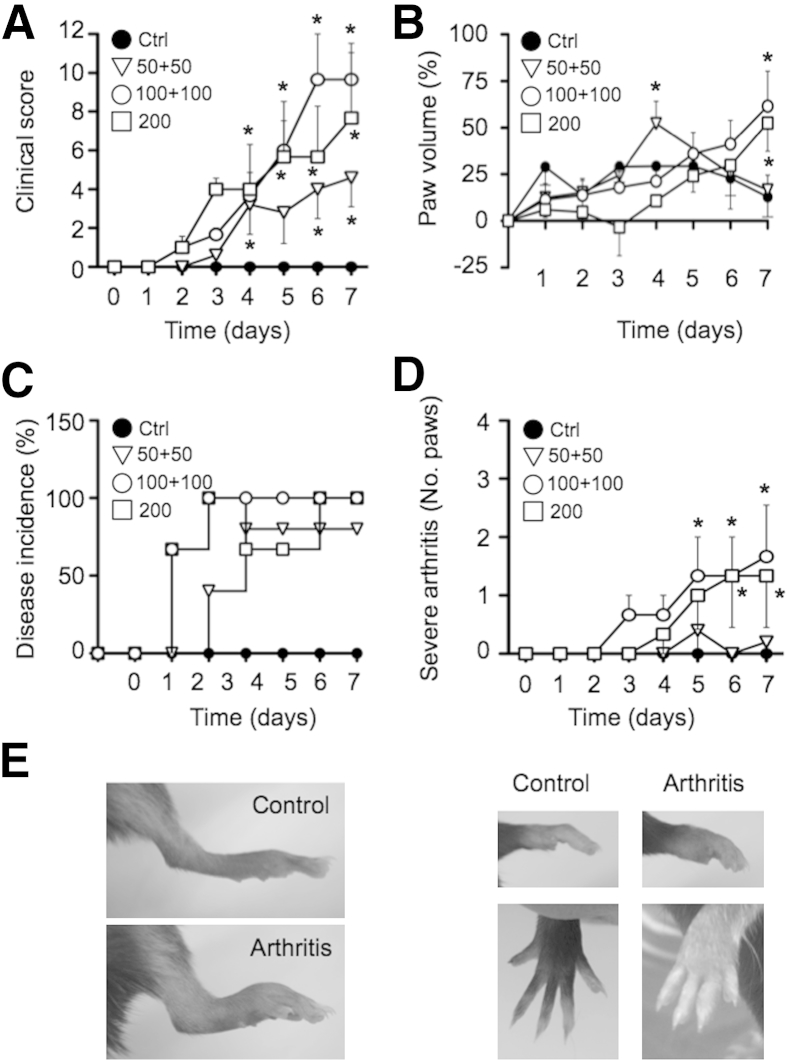
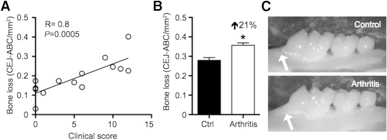
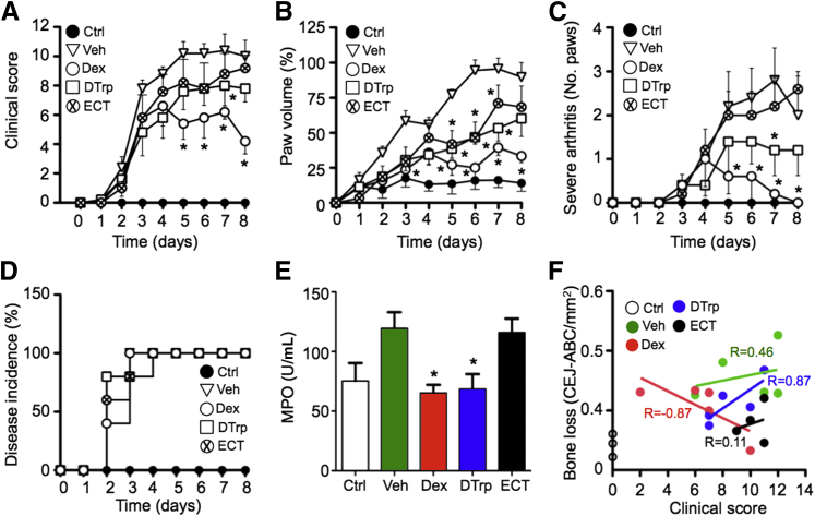
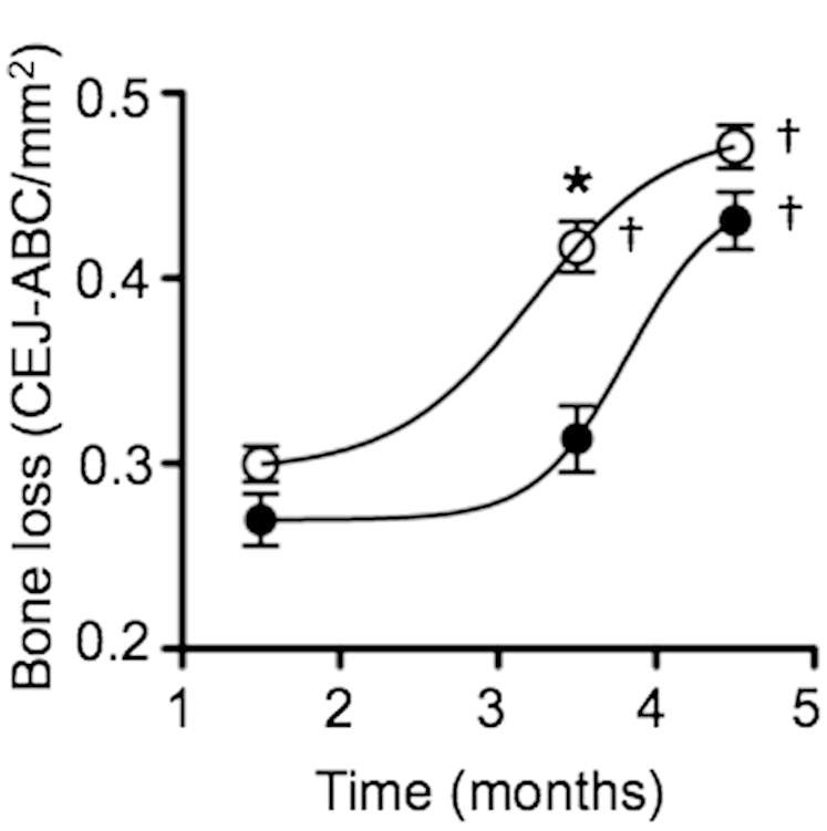
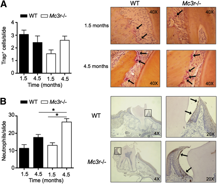
Because there is clinical evidence for an association between periodontal disease and rheumatoid arthritis, it is important to develop suitable experimental models to explore pathogenic mechanisms and therapeutic opportunities. The K/BxN serum model of inflammatory arthritis was applied using distinct protocols, and modulation of joint disruption afforded by dexamethasone and calcitonin was established in comparison to the melanocortin (MC) receptor agonist DTrp(8)-γ-melanocyte stimulating hormone (MSH; DTrp). Wild-type and MC receptor type 3 (MC3)-null mice of different ages were also used. There was significant association between severity of joint disease, induced with distinct protocols and volumes of the arthritogenic K/BxN serum, and periodontal bone damage. Therapeutic treatment with 10 μg dexamethasone, 30 ng elcatonin, and 20 μg DTrp per mouse revealed unique and distinctive pharmacological properties, with only DTrp protecting both joint and periodontal tissue. Further analyses in nonarthritic animals revealed higher susceptibility to periodontal bone loss in Mc3r(-/-) compared with wild-type mice, with significant exacerbation at 14 weeks of age. These data reveal novel protective properties of endogenous MC3 on periodontal status in health and disease and indicate that MC3 activation could lead to the development of a new genus of anti-arthritic bone-sparing therapeutics.


Figures

Figure 1
Figure 1
Comparison of three protocols for the K/BxN serum transfer arthritis model. Arthritis was induced by the i.p. injection of serum from K/BxN arthritic mice using three different protocols: 50 + 50 (two injections of 50 μL on days 0 and 2); 100 + 100 (two injections of 100 μL on days 0 and 2); and 200 (one single injection on day 0). Clinical score (A), paw volume (B), and disease incidence (C) were recorded for 7 days. D: The number of paws per mouse that reached the maximum score (3). E: Representative images of ankle, wrist, and digit swelling. Data are the means ± SEM of four to six mice per group. P < 0.05, two-way analysis of variance, followed by Bonferroni multiple-comparison test. Ctrl, control.
Figure 2
Figure 2
Correlation of arthritis with alveolar bone loss in the K/BxN serum transfer model. Alveolar bone loss was evaluated at day 7 in the palatal aspect of the first upper molar of the right hemimaxillae. A: Correlation between alveolar bone loss and clinical score on mice studied in the protocol comparison experiment (Figure 1) analyzed by Pearson correlation test (n = 14). B: Overall increase in alveolar bone loss in arthritic mice (pooled data from all mice, means ± SEM, n = 14) compared with control (Ctrl) mice, analyzed by t-test. P < 0.05. C: Representative photographs of the maxillae, showing evidence of alveolar bone loss (arrows).
Figure 3
Figure 3
Effect of dexamethasone, DTrp, and ECT in arthritis and alveolar bone loss. Arthritis was induced using the 100 + 100 protocol (100 μL of serum on days 0 and 2) and monitored by daily recording the clinical score (A), paw volume (B), severity (number of paws reaching the maximum score) (C), and disease incidence (D). E: MPO activity was measured in the left hemimaxillae at day 8. F: Alveolar bone loss was analyzed in the right hemimaxillae at day 8 and correlated with clinical score recorded that day. Drugs were administered i.p. once daily: Dex, 10 μg per mouse; DTrp, 20 μg per mouse; ECT, 30 ng per mouse; and vehicle PBS (Veh). Nonarthritic mice were included as controls (Ctrl). Data are the means ± SEM of five to six mice per group. Data were analyzed by two-way analysis of variance, followed by a Bonferroni multiple-comparison test (AC), one-way analysis of variance, followed by a Bonferroni multiple-comparison test (E), and Pearson correlation test (F). P < 0.05.
Figure 4
Figure 4
Arthritis and alveolar bone loss in MC3-deficient mice. Arthritis was induced in C57BL/6J WT mice (black) and melanocortin receptor 3–deficient mice (Mc3r−/−) (white) using the 100 + 100 protocol (100 μL of serum on days 0 and 2). Disease was monitored by daily recording of the clinical score (A), paw volume (B), and disease severity (number of paws reaching the maximum score) (C). Alveolar bone loss was analyzed in the right hemimaxillae on the last day of the experiment (day 8). Data are the means ± SEM of five mice per group. Statistical analyses were performed by two-way analysis of variance, followed by a Bonferroni multiple-comparison test (AC), and one-way analysis of variance, followed by a Newman-Keuls multiple-comparison test versus WT-control (Ctrl; D). P < 0.05.
Figure 5
Figure 5
Impact of aging on alveolar bone loss in MC3-deficient mice. Alveolar bone loss was evaluated in the right hemimaxillae in mice from different ages (1.5, 3.5, and 4.5 months old) in both C57BL/6J WT mice (white circles) and melanocortin receptor 3–deficient mice (Mc3r−/−) (black circles). Data are the means ± SEM of 6 to 17 mice. Data were analyzed by two-way analysis of variance, followed by a Bonferroni multiple-comparison test. P < 0.05 versus 1.5 months; P < 0.05 for WT versus Mc3r−/−.
Figure 6
Figure 6
Analysis of osteoclasts and neutrophils in gingival tissues. The left hemimaxillae were used for histological evaluation of osteoclast activity by Trap staining and neutrophil infiltration. A: The number of osteoclasts on the cervical area of the first molar. Representative images of Trap+ cells (arrows). B: Sections were stained for neutrophil elastase as a marker of neutrophils. Representative images of 1.5-month-old mice. Arrows designate neutrophils. Data are the means ± SEM of two to four mice. Data were analyzed by one-way analysis of variance, followed by a Bonferroni multiple-comparison test. P < 0.05.

Comment in

Similar articles

Cited by

References

    1. McInnes I.B., Schett G. The pathogenesis of rheumatoid arthritis. N Engl J Med. 2011;365:2205–2219. - PubMed
    1. Boissier M.C., Semerano L., Challal S., Saidenberg-Kermanac'h N., Falgarone G. Rheumatoid arthritis: from autoimmunity to synovitis and joint destruction. J Autoimmun. 2012;39:222–228. - PubMed
    1. Taylor P.C. Developing anti-TNF and biologic agents. Rheumatology (Oxford) 2011;50:1351–1353. - PubMed
    1. Gantz I., Fong T.M. The melanocortin system. Am J Physiol Endocrinol Metab. 2003;284:E468–E474. - PubMed
    1. Catania A., Lonati C., Sordi A., Carlin A., Leonardi P., Gatti S. The melanocortin system in control of inflammation. ScientificWorldJournal. 2010;10:1840–1853. - PMC - PubMed

Publication types

Substances