. 2014 Jul 22;111(29):10580-5.
doi: 10.1073/pnas.1401591111. Epub 2014 Jun 16.

An uncoupling channel within the c-subunit ring of the F1FO ATP synthase is the mitochondrial permeability transition pore

Affiliations

An uncoupling channel within the c-subunit ring of the F1FO ATP synthase is the mitochondrial permeability transition pore

Kambiz N Alavian et al. Proc Natl Acad Sci U S A. .

Abstract

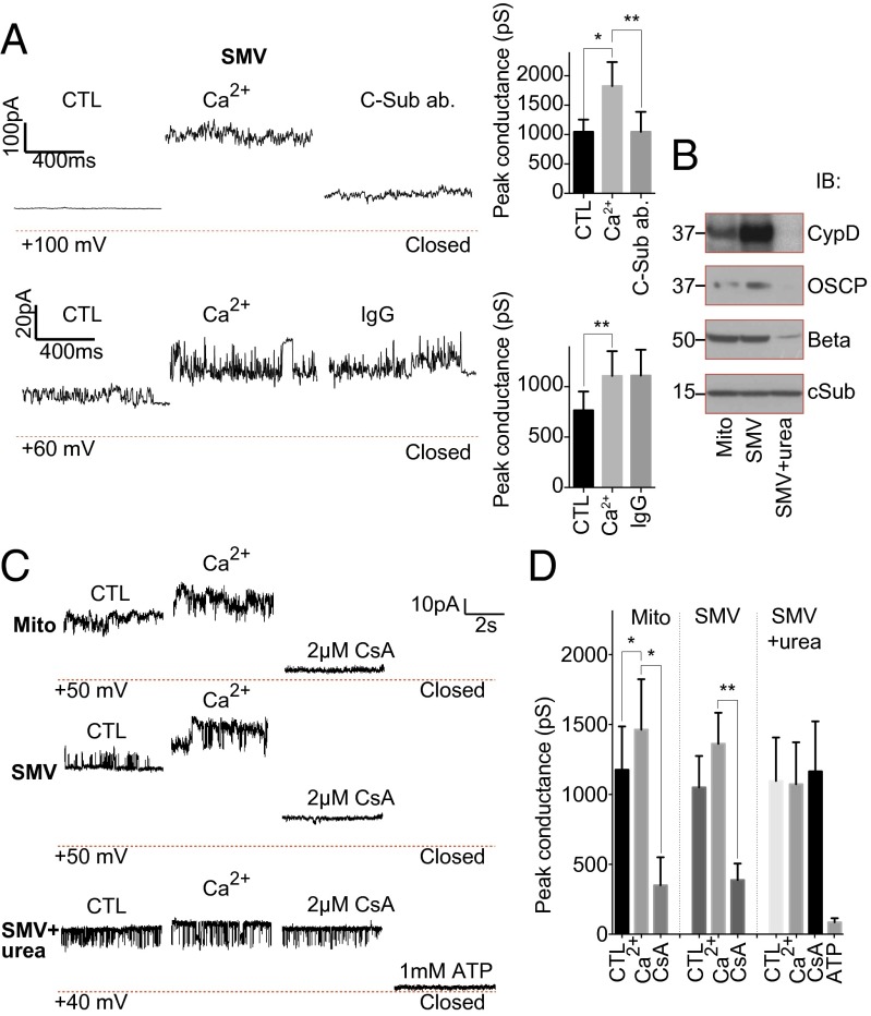
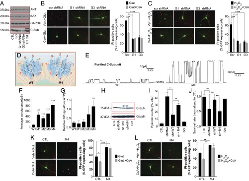
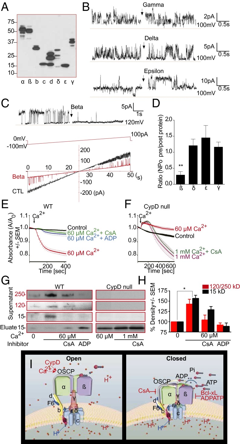
Mitochondria maintain tight regulation of inner mitochondrial membrane (IMM) permeability to sustain ATP production. Stressful events cause cellular calcium (Ca(2+)) dysregulation followed by rapid loss of IMM potential known as permeability transition (PT), which produces osmotic shifts, metabolic dysfunction, and cell death. The molecular identity of the mitochondrial PT pore (mPTP) was previously unknown. We show that the purified reconstituted c-subunit ring of the FO of the F1FO ATP synthase forms a voltage-sensitive channel, the persistent opening of which leads to rapid and uncontrolled depolarization of the IMM in cells. Prolonged high matrix Ca(2+) enlarges the c-subunit ring and unhooks it from cyclophilin D/cyclosporine A binding sites in the ATP synthase F1, providing a mechanism for mPTP opening. In contrast, recombinant F1 beta-subunit applied exogenously to the purified c-subunit enhances the probability of pore closure. Depletion of the c-subunit attenuates Ca(2+)-induced IMM depolarization and inhibits Ca(2+) and reactive oxygen species-induced cell death whereas increasing the expression or single-channel conductance of the c-subunit sensitizes to death. We conclude that a highly regulated c-subunit leak channel is a candidate for the mPTP. Beyond cell death, these findings also imply that increasing the probability of c-subunit channel closure in a healthy cell will enhance IMM coupling and increase cellular metabolic efficiency.

Keywords: apoptosis; excitotoxicity; ion channel; metabolism; necrosis.


Conflict of interest statement

The authors declare no conflict of interest.

Figures

Fig. 1.
Fig. 1.
ATP synthase c-subunit ring forms a large conductance ion channel. (A, Upper) Excised proteoliposome patch-clamp recording of purified c-subunit protein. (Left) Amplitude histogram shows conductances of ∼227 pS, 1,450 pS, and 1,680 pS; C, closed; O1–O3, open. (Right) Amplitude histograms of six random recordings taken after depolarization of the patch. The histogram from the Left is indicated in red. C is closed; O1–O3 mark the open levels at conductances of ∼100–300 pS, ∼1,500 pS, and ∼1,800–2,000 pS. Conductance levels of ∼500–750 pS are also seen but not designated in the figure. (B) Representative (∼600 pS) excised proteoliposome patch-clamp recording of purified c-subunit protein during continuous ramp voltage clamp from −100 mV to +100 mV before and after 1 mM ATP. (C) Representative patch-clamp recordings of purified c-subunit protein before and after exposure to anti–pan-ATP5G1/2/3 (C-Sub ab.), control antibody (IgG), or 2 µM CsA. (D) Quantification of peak conductance before and after exposure to anti-ATP5G1/2/3 (n = 10, **P = 0.0022), control IgG (n = 3), 100 µM Ca2+ (n = 3), or 2 µM CsA (n = 4). (E) Representative patch-clamp recordings of purified ATP synthase reconstituted into liposomes before and after addition of (in sequence) recombinant CypD protein (5 µg/mL final), 100 µM CaCl2, and 5 µM CsA. (F) Group data for recordings represented in E (n = 12; *P = 0.0228, **P = 0.00391). Closed state of channel activity indicated by the dotted red line.
Fig. 2.
Fig. 2.
The c-subunit large conductance ion channel is present in mitochondria. (A) Representative (∼1,900 pS, Upper; ∼1,000 pS, Lower) SMV patch-clamp recordings before and after exposure to 100 µM Ca2+ followed by anti-ATP5G1/2/3 (C-sub ab.) or control antibody (IgG). Bar graphs show quantification of peak conductances (Upper, N = 8, *P = 0.0194, **P = 0.0033; Lower, N = 5, **P = 0.0256). (B) SDS immunoblot of mitochondria, SMVs, and urea-treated SMVs. C-sub ab is to ATP5G1. (C) Representative patch-clamp recordings of whole-liver mitochondria, SMVs, and urea-exposed SMVs before and after treatment with Ca2+ then CsA and ATP (Bottom trace). (D) Peak conductance in response to Ca2+, CsA, and ATP (n = 4 Mito, 5 SMV, 10 urea-treated SMV, 5 ATP; *P = 0.0045, **P < 0.0001). Closed (0 pA) indicated by the dotted red line.
Fig. 3.
Fig. 3.
The c-subunit ring regulates Ca2+-induced IMM uncoupling. (A) Schematic of cysteine pair labeling of two adjacent c-subunit monomers in the open and closed position. Binding by FlAsh and subsequent fluorescence occur when cysteine pairs move together. (B) Representative TMRE, FlAsh fluorescence, and merged image in HEK 293T cells. (C) Time course of FlAsh fluorescence intensity in HEK 293T cells 72 h after transfection (CC-CC indicates two cysteine pairs, and CC indicates one cysteine pair). Cells were pretreated with vehicle or 2 µM CsA for 1 h and exposed to 2 µM ionomycin (Ion., arrow). (D) Group data for FlAsh fluorescence intensity (n = 12 data points and ≥3 independent experiments per condition; *P < 0.05, **P < 0.01, ***P < 0.0001, N.S., not significant). (E) Time course of calcein fluorescence intensity before and after addition of 1 µM (for CsA experiment) or 4 µM ionomycin to HEK 293T cells treated with 5 µM CsA or transfected with the indicated shRNA constructs (Scr, scrambled). (F) Group data for experiment in E at 20 min after addition of ionomycin (n = 3 cells, 16 cells, 13 cells, 18 cells for CsA (1 culture), scrambled, ATP5G3, and ATP5G1 shRNA (three independent cultures) (***P < 0.0001). (Scale bar: 10 μm.)
Fig. 4.
Fig. 4.
Altering c-subunit expression or packing regulates IMM conductance and sensitivity to excitotoxic and ROS-mediated cell death. (A) Immunoblot using the indicated antibodies. (B and C) Images and group data of propidium iodide (PI) staining of cultured hippocampal neurons at 24 h after 30-min exposure to 100 µM glutamate or 20 µM H2O2 with or without 0.5 µM CsA (n ≥ 3 independent cultures, 56–65 micrographs of each condition; G1, ATP5G1; G3, ATP5G3). (D) Schematic of the WT and M4 c-subunit rings. Increased channel conductance of M4 mutant suggests decreased packing and a larger central pore due to the Gly-Val mutation. (E) Representative patch clamp recordings of liposomes reconstituted with WT or M4 c-subunit. (F) Peak single channel conductance for liposome recordings with the indicated protein (WT, n = 38; M1, n = 7; M2, n = 17; M3, n = 7 M4, n = 22; ***P < 0.0001 for WT vs. M1–M4). (G) Normalized NPO before and after addition of 1 mM ATP to patches with the indicated protein (WT, n = 21; M1, n = 3; M2, n = 6; M3, n = 5; M4, n = 8; ***P = 0.0009 comparing WT to M4 ATP response, NPO, number of channels multiplied times the probability of opening of each channel). (H) Immunoblot using the indicated antibodies. The uppermost bands labeled with anti–c-subunit antibody represent the myc/FLAG tagged overexpressed protein. The lower bands have lost one of the tags (blot is representative of >5 blots). (I and J) Percent pyknotic nuclei (I) and cell survival (MTT assay) (J), measured in HEK 293T cells expressing the indicated constructs (n = 2 experiments with three coverslips, two fields per coverslip per condition (**P < 0.01, ***P < 0.0001). Measurements were made 24 h after switching to galactose-containing, glucose-free medium. (K and L) Images and group data of PI staining of hippocampal neurons at 18 h after 30-min exposure to 100 µM glutamate or 20 µM H2O2 with vehicle or 0.5 µM CsA (n = 3 independent cultures, 38–53 micrographs of each condition. (Scale bars: B, C, K, and L, 20 µm.) *P < 0.05, **P < 0.01, ***P < 0.001, ****P < 0.0001.
Fig. 5.
Fig. 5.
Structural rearrangements of ATP synthase unmask the c-subunit pore. (A) Immunoblot using antibody against mammalian myc/FLAG-tagged human ATP synthase subunit proteins (blot representative of 3 blots). (B and C) Representative proteoliposome patch clamp recordings of purified c-subunit. Purified gamma, delta, epsilon or beta-subunit (50 ng/µL recording solution) were added (arrow). (C, Lower) Ramp voltage recording. (D) NPO ratio after/before addition of indicated ATP synthase subunits (n = 5, **P = 0.0036). (E and F) Ca2+-induced IMM swelling assay of WT heart or CypD KO mitochondria in 200 nM CsA or 0.5 mM ADP. Plotted is ratio of absorbance at 540 nm (A) after/before Ca2+ (n ≥ 3 each). (G) Immunoblots (ATP5G-com or ATP5G1) of the supernatants and eluates after ATP synthase IP from WT and CypD KO mitochondrial lysates, as in E and F (representative of ≥4 blots). (H) Quantification of 15-kDa band and 120 plus 250 kDa oligomer band density in gels represented in G (n ≥ 3 for all bands, *P = 0.0331 for 120 plus 250 kDa and 0.0417 for 15 kDa compared with control). (I) Schematic of regulation of PT (c-subunit) pore by ATP synthase F1. (Left) CypD and Ca2+ expand the c-subunit ring and open the pore by releasing F1. (Right) CsA, Bcl-xL, and ADP/ATP close the c-subunit pore by positioning beta-subunit over its mouth.

Comment in

References

    1. Petronilli V, Szabò I, Zoratti M. The inner mitochondrial membrane contains ion-conducting channels similar to those found in bacteria. FEBS Lett. 1989;259(1):137–143. - PubMed
    1. Antonenko YN, Smith D, Kinnally KW, Tedeschi H. Single-channel activity induced in mitoplasts by alkaline pH. Biochim Biophys Acta. 1994;1194(2):247–254. - PubMed
    1. Bernardi P, et al. Modulation of the mitochondrial permeability transition pore: Effect of protons and divalent cations. J Biol Chem. 1992;267(5):2934–2939. - PubMed
    1. Bernardi P. Modulation of the mitochondrial cyclosporin A-sensitive permeability transition pore by the proton electrochemical gradient: Evidence that the pore can be opened by membrane depolarization. J Biol Chem. 1992;267(13):8834–8839. - PubMed
    1. Jonas EA, Buchanan J, Kaczmarek LK. Prolonged activation of mitochondrial conductances during synaptic transmission. Science. 1999;286(5443):1347–1350. - PubMed

Publication types

MeSH terms