. 2014 Jul 1;111(26):9633-8.
doi: 10.1073/pnas.1404651111. Epub 2014 Jun 16.

Transcellular degradation of axonal mitochondria

Affiliations

Transcellular degradation of axonal mitochondria

Chung-ha O Davis et al. Proc Natl Acad Sci U S A. .

Abstract

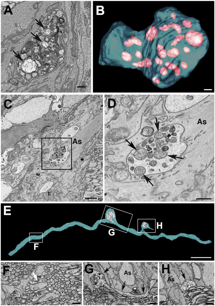
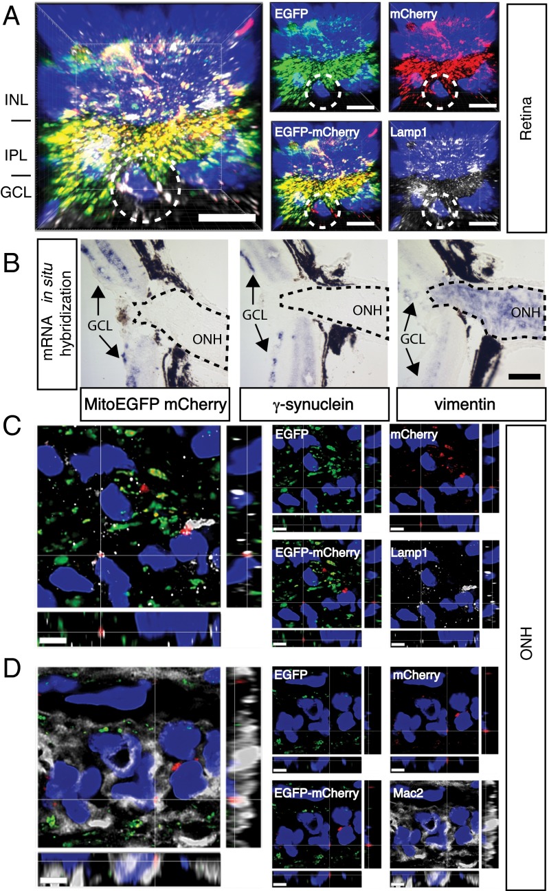
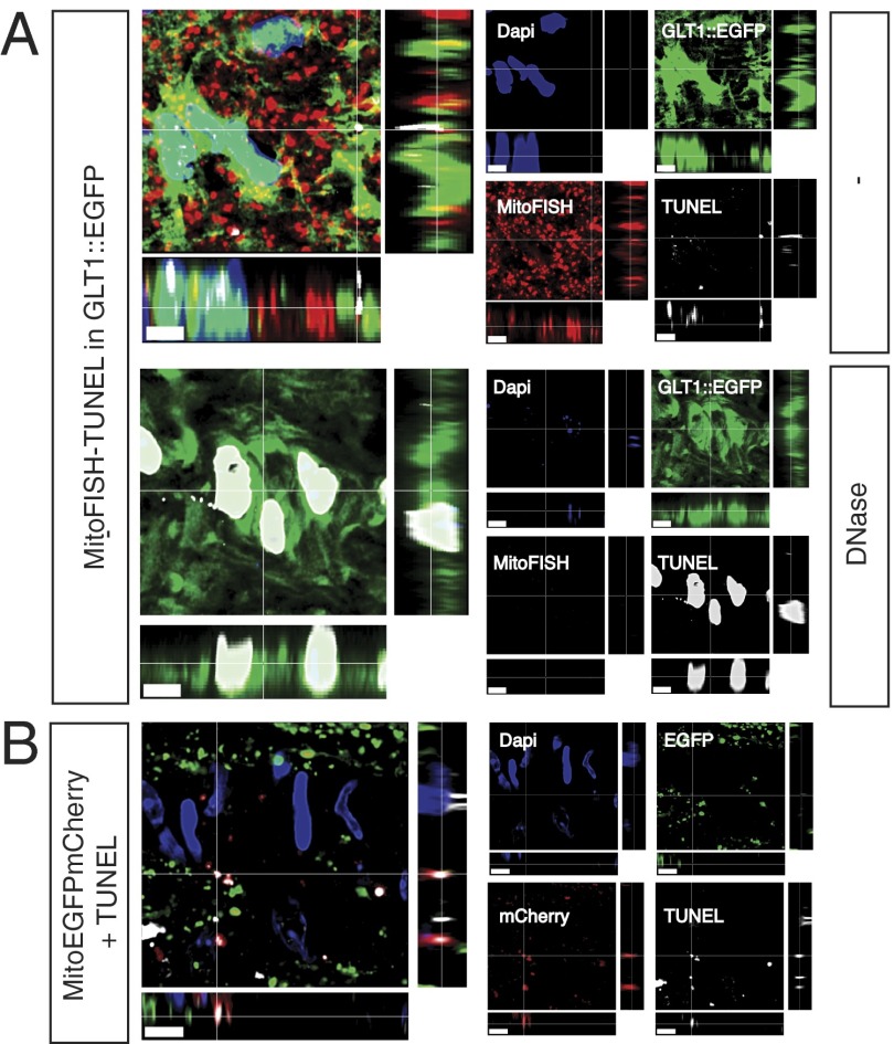
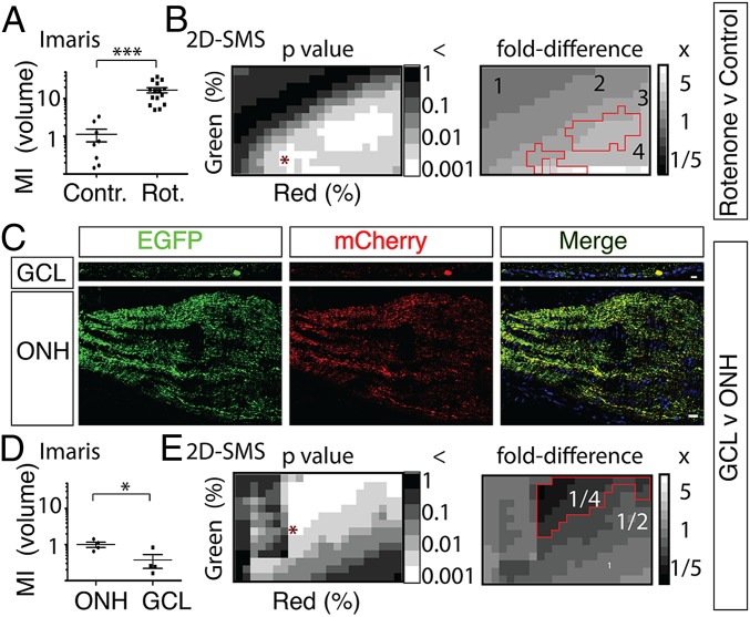
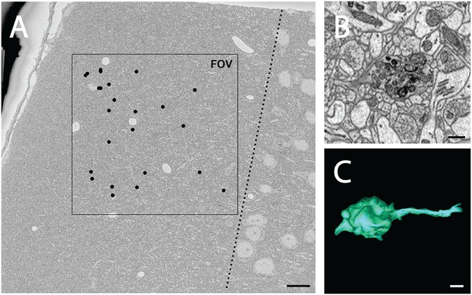
It is generally accepted that healthy cells degrade their own mitochondria. Here, we report that retinal ganglion cell axons of WT mice shed mitochondria at the optic nerve head (ONH), and that these mitochondria are internalized and degraded by adjacent astrocytes. EM demonstrates that mitochondria are shed through formation of large protrusions that originate from otherwise healthy axons. A virally introduced tandem fluorophore protein reporter of acidified mitochondria reveals that acidified axonal mitochondria originating from the retinal ganglion cell are associated with lysosomes within columns of astrocytes in the ONH. According to this reporter, a greater proportion of retinal ganglion cell mitochondria are degraded at the ONH than in the ganglion cell soma. Consistently, analyses of degrading DNA reveal extensive mtDNA degradation within the optic nerve astrocytes, some of which comes from retinal ganglion cell axons. Together, these results demonstrate that surprisingly large proportions of retinal ganglion cell axonal mitochondria are normally degraded by the astrocytes of the ONH. This transcellular degradation of mitochondria, or transmitophagy, likely occurs elsewhere in the CNS, because structurally similar accumulations of degrading mitochondria are also found along neurites in superficial layers of the cerebral cortex. Thus, the general assumption that neurons or other cells necessarily degrade their own mitochondria should be reconsidered.

Keywords: mitophagy; phagocytosis.


Conflict of interest statement

The authors declare no conflict of interest.

Figures

Fig. 1.
Fig. 1.
Evulsions of retinal ganglion cell axons within the ONH contain mitochondria. An SBEM single section (A) and serial reconstruction (B) of axonal evulsion within the ONH of a 3-mo-old C57BL/6J mouse containing morphologically distinct mitochondria (arrows in A and pink volumes in B). Transmission EM (C) and enlarged view of the boxed area (D) showing mitochondria with normal morphology (arrows) and mitochondria remnants within the same evulsion. (E) SBEM-based reconstruction of a single axon displaying two protrusions (boxes G and H). (FH) Sections through the areas boxed in E. The white arrow in F points to close apposition between axons without intervening glia, and the black arrows in G and H point to direct contacts between the axon and astrocyte processes (As). (Scale bars: A, B, and D, 0.5 μm; C, 1 μm; E, 5 μm; FH, 2 μm.)
Fig. 2.
Fig. 2.
AAV2::MitoEGFPmCherry infection of the retina demonstrates that some retinal ganglion cell mitochondria are degraded within ONH astrocytes. (A) mCherry-ONLY puncta are within the retinal GCL of AAV2:MitoEGFPmCherry-infected retinas and colocalize with the lysosomal marker Lamp1. Circles surround a GCL cell with prominent mCherry-ONLY puncta. INL, inner nuclear layer; IPL, inner plexiform layer. (B) AAV::MitoEGFPmCherry mRNA is found only in the retina, including the GCL, where ganglion cells are labeled by γ-synuclein mRNA, but not in the ONH, where astrocytes are labeled by vimentin mRNA. Dotted traces demarcate the ONH. (C) Within the ONH, common mCherry-ONLY puncta overlap with or are surrounded by the lysosomal marker Lamp1. A single mCherry-ONLY punctum surrounded by multiple vesicles containing Lamp1 is shown in the xyz projection. (D) Within the ONH MTZ, the mCherry-ONLY puncta localize largely separate from axon bundles and within the glial columns, and colocalize with the MTZ astrocyte cytoplasmic marker Mac2. Single mCherry-ONLY punctum surrounded by Mac2-labeled cytoplasm is shown in the xyz projection. All images show nuclei labeled by DAPI as a reference. (Scale bars: A, C, and D, 5 μm; B, 100 μm.)
Fig. 3.
Fig. 3.
Combination of TUNEL and MitoFISH identifies mitochondria degraded by astrocytes within the ONH and confirms that some of these degrading mitochondria are derived from axons. (A) In sections of ONH where astrocytes are labeled by a GLT1::EGFP BAC transgene and mtDNA is detected by MitoFISH, TUNEL labels degrading mtDNA within astrocyte soma and processes; after DNase treatment, TUNEL only labels nuclei remnants. (B) In the ONH of C57BL/6J mice whose retinal ganglion mitochondria are labeled by the AAV2::MitoEGFPmCherry transgene, the TUNEL signal colocalizes with the mCherry-ONLY puncta. (Scale bars: 5 μm.)
Fig. 4.
Fig. 4.
Three-dimensional and 2D quantitative assays based on the Cherry-ONLY signal after AAV2::MitoEGFPmCherry intraocular injection demonstrate that a higher proportion of retinal ganglion cell mitochondria are degraded in the ONH relative to the GCL. (A) Intravitreal injection of 0.5 μL of 3.1 mM rotenone (Rot.) produces a 16-fold increase (***P < 0.001, Student t test) in the MI by volume [MI (volume)] within the ONH, as measured by subjectively set volume thresholding in Imaris software. Representative images are shown in Fig. S4A. Contr., control. (B) Two-dimensional SMS, which analyses 2D images at multiple segmentation values across two color channels, also shows that rotenone produces significant increases in MIarea, which range from 2.3- to 6.4-fold increases depending on the segmentation used but average a 3.4 ± 0.9-fold increase at the P < 0.001 significance level (Student t test). Representative images binarized at the segmentation pair denoted by an asterisk in P value distribution are shown in Fig. S4C. MIarea P values and fold differences for a scrambled control are shown in Fig. S4E. (C) Representative images of MitoEGFPmCherry labeling in the GCL and ONH from the same eye. (Scale bar: 20 μm.) (D) MI (volume) is lower (*P < 0.05, Student t test) in the GCL relative to the ONH, as determined by comparing ONH sections (n = 4) with retina flat-mount images (n = 4) of one mouse using subjective thresholding in Imaris software. (E) Comparing sections of the GCL and ONH from multiple animals (n = 10) using 2D-SMS also shows significantly lower MIarea in the GCL relative to the ONH, with MI fold differences ranging from 0.03 to 0.30 depending on the segmentation used and averaging a 0.22 ± 0.16-fold difference at the P < 0.001 level (paired Student t test). Representative images of GCL and ONH binarized at the segmentation pair denoted by an asterisk in P value distribution are shown in Fig. S4D. MIarea P values and fold differences for a scrambled control are shown in Fig. S4F.
Fig. 5.
Fig. 5.
Protrusions containing degrading mitochondria are found in superficial layers of cerebral cortex of a young WT mouse. (A) Low-magnification view of a region of cortical layer 1 of a 3-mo-old C57BL/6J mouse imaged with SBEM, where black dots indicate the locations of protrusions found within volume. Total volume dimensions were 200 μm × 200 μm and 32.8 μm in depth. The dotted line indicates the boundary between layers 1 and 2. (B) Higher magnification views of a protrusion reveal morphology similar to that seen in the ONH, consisting of large, round, membrane-bound bodies filled with debris particles, including mitochondrial fragments. (C) Three-dimensional reconstruction of the same protrusion. (Scale bars: A, 15 μm; B and C, 0.5 μm.)

Comment in

References

    1. Scarpulla RC, Vega RB, Kelly DP. Transcriptional integration of mitochondrial biogenesis. Trends Endocrinol Metab. 2012;23(9):459–466. - PMC - PubMed
    1. Ashrafi G, Schwarz TL. The pathways of mitophagy for quality control and clearance of mitochondria. Cell Death Differ. 2013;20(1):31–42. - PMC - PubMed
    1. Youle RJ, van der Bliek AM. Mitochondrial fission, fusion, and stress. Science. 2012;337(6098):1062–1065. - PMC - PubMed
    1. Nguyen JV, et al. Myelination transition zone astrocytes are constitutively phagocytic and have synuclein dependent reactivity in glaucoma. Proc Natl Acad Sci USA. 2011;108(3):1176–1181. - PMC - PubMed
    1. Pankiv S, et al. p62/SQSTM1 binds directly to Atg8/LC3 to facilitate degradation of ubiquitinated protein aggregates by autophagy. J Biol Chem. 2007;282(33):24131–24145. - PubMed

Publication types

Substances