. 2014 Jul 1;111(26):9503-8.
doi: 10.1073/pnas.1408638111. Epub 2014 Jun 16.

Molecular ties between the cell cycle and differentiation in embryonic stem cells

Affiliations

Molecular ties between the cell cycle and differentiation in embryonic stem cells

Victor C Li et al. Proc Natl Acad Sci U S A. .

Abstract

Attainment of the differentiated state during the final stages of somatic cell differentiation is closely tied to cell cycle progression. Much less is known about the role of the cell cycle at very early stages of embryonic development. Here, we show that molecular pathways involving the cell cycle can be engineered to strongly affect embryonic stem cell differentiation at early stages in vitro. Strategies based on perturbing these pathways can shorten the rate and simplify the lineage path of ES differentiation. These results make it likely that pathways involving cell proliferation intersect at various points with pathways that regulate cell lineages in embryos and demonstrate that this knowledge can be used profitably to guide the path and effectiveness of cell differentiation of pluripotent cells.

Keywords: differentiation modeling; guided differentiation; proliferation control; systems biology.


Conflict of interest statement

The authors declare no conflict of interest.

Figures
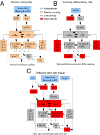
Fig. 1.
Fig. 1.
Suggested summary of the proliferation/differentiation network adapted for ES cells. (A) Reconstructed summary of main network components and interactions in normal somatic cycling cells. Extracellular growth factors (GFs) either purified or in serum activate downstream signaling pathways (PI3K, MAPK), which then trigger a transcriptional activation (Myc, E2F) that drives the core cell cycle machinery (Cyclins and Cyclin-dependent kinases). For a full explanation and justification of the summary, see Fig. S1. The cellular behavior associated with this model is normal oscillatory cycling. (B) Changes to the network that occur during terminal division arrest and differentiation. Cells switch to insulin signaling for survival and growth and shut down cycling activity. Terminal transcription factors become fully active, leading to complete differentiation. (C) Composite adaptation of network for ES cells. ES cells are normally maintained by LIF and high serum or LIF and Bmp4. This hyperactivates PI3K, Myc, E2F, CDK2, and Id family activities. Meanwhile, MAPK, CDK4, p16 family, p21 family, and Rb family activities are highly suppressed. This leads to an ultrarapid proliferation and short G1 phase.
Fig. 2.
Fig. 2.
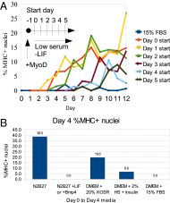
Growth factor/serum reduction drives a direct terminal skeletal muscle differentiation program. (A) Differentiation time course of ES cells to skeletal muscle when exposed to low serum vs. high serum conditions. MyoD overexpression was initially induced with tamoxifen starting at day −1 for 24 h under ES conditions. LIF was removed at day 0. Low serum was then initiated at different starting times beginning with day 0. Cells were then fixed and immunostained for MHC expression. Differentiating cells in high serum were continuously split to prevent overgrowth. (B) A comparison of differentiation efficiency generated by two types of defined media (N2B27) and 20% KOSR, low serum media [2% horse serum (HS) + insulin], and high serum media (15% FBS). Cells were removed from standard ES media (LIF and serum) and incubated in the specified media starting from day 0 to day 4. Continuation of LIF (1000U/mL) or addition of Bmp4 (10 ng/mL) results in a strong block to muscle differentiation.
Fig. 3.
Fig. 3.
Growth factor/serum withdrawal drives a condensed gene expression program of early Pax3 expression and subsequent MyoG expression but does not affect kinetics of Oct4 or Nanog loss. Gene expression time courses of differentiation over 7 d. In this experiment, low serum was initiated at day 0, 1 d after induction of MyoD expression in ES media, and maintained over the course of differentiation. The rate of decline of Oct4 and Nanog mRNA levels are not strongly affected by cell cycle inhibition. In the muscle regulatory hierarchy, only Pax3 and MyoG are strongly up-regulated. Up-regulation of additional terminal markers of skeletal muscle terminal differentiation can be found in Fig. S3. Ct values were normalized to glyceraldehyde 3-phosphate dehydrogenase (GADPH). Error values reflect SEM (n = 3).
Fig. 4.
Fig. 4.
Specific cell cycle inhibitors have stage and condition-specific effects on Pax3 and MyoG expression. Time courses of differentiation were run for 7 d, similarly to the previous growth factor withdrawal experiments. MyoD was induced at day −1, and LIF was removed at day 0. Cells were kept in either high serum (15% FBS) or low serum [2% horse serum (HS) plus insulin] media throughout the time course. Drug treatments were applied continuously at the designated concentrations for the entire duration of the time course, with daily media change. mRNA expression levels were measured by quantitative RT-PCR. Pax3 was measured on day 3 (because it is up-regulated early), and MyoG was measured on day 7 (because it is up-regulated late). All measurements are relative to a DMSO control for the specific condition (i.e., low serum values are still higher than in high serum). L, lethal (drug had strong antisurvival effects, so no data were collected). Error bars indicate SEM (n = 2).
Fig. 5.
Fig. 5.
In an unguided or heterogeneous differentiation setting, growth factor/serum withdrawal up-regulates the expression of numerous genes associated with differentiated cell types. Gene expression time courses of unguided (without MyoD) differentiation after release from ES cell media at day 0. Low or high serum was applied at day 0 for the full time course. Up-regulated factors include Dll1, Sox6, GATA1, PPARγ, Sox9, Runx2, Mitf, Sox17, and Nkx2.2 (continued in Fig. S4). These genes are involved in the differentiation of multiple cell types, including neurons, chondrocytes, erythrocytes, adipocytes, cardiomyocytes, osteoblasts, melanocytes, and β cells. Error values indicate SEM (n = 2).
Fig. 6.
Fig. 6.
Use of network-based cell cycle manipulation may provide an alternative strategy to generating terminal cell types that is more efficient than recapitulating embryogenesis. (Upper) Analytical scheme of how cell cycle states drive activation of a terminal transcription factor. In our MyoD overexpression system, we propose that transitions from the three states of ES to somatic cycling to a terminally differentiated state influence the subsequent activation of MyoD and hence the progression of differentiation. The first transition is correlated with Pax3 activation, and the second transition is correlated with MyoG activation. Accelerating the cell cycle transitions accelerates the differentiation process. (Lower) Current strategies of differentiating ES cells into cells involves growth-factor based recapitulation of embryogenesis. The alternative strategy suggested in this paper (and shown in the case of skeletal muscle) is to artificially induce differentiation by accelerating cell cycle inhibition combined with addition of a terminal transcription factor. Because ES cells are susceptible to cell cycle-related pathways, this can lead to a faster, more efficient differentiation.

References

    1. Muñoz-Alonso M, León J. G1 phase control and cell differentiation. In: Boonstra J, editor. G1 Phase Progression. Georgetown, TX: Landes Bioscience; 2003. pp. 236–264.
    1. Budirahardja Y, Gönczy P. Coupling the cell cycle to development. Development. 2009;136(17):2861–2872. - PubMed
    1. Hindley C, Philpott A. Co-ordination of cell cycle and differentiation in the developing nervous system. Biochem J. 2012;444(3):375–382. - PMC - PubMed
    1. Li VC, Ballabeni A, Kirschner MW. Gap 1 phase length and mouse embryonic stem cell self-renewal. Proc Natl Acad Sci USA. 2012;109(31):12550–12555. - PMC - PubMed
    1. Thoma EC, Maurus K, Wagner TU, Schartl M. Parallel differentiation of embryonic stem cells into different cell types by a single gene-based differentiation system. Cell Reprogram. 2012;14(2):106–111. - PubMed

Publication types

MeSH terms