

Role of diacylglycerol activation of PKCθ in lipid-induced muscle insulin resistance in humans
- PMID: 24979806
- PMCID: PMC4084449
- DOI: 10.1073/pnas.1409229111
Role of diacylglycerol activation of PKCθ in lipid-induced muscle insulin resistance in humans
Abstract
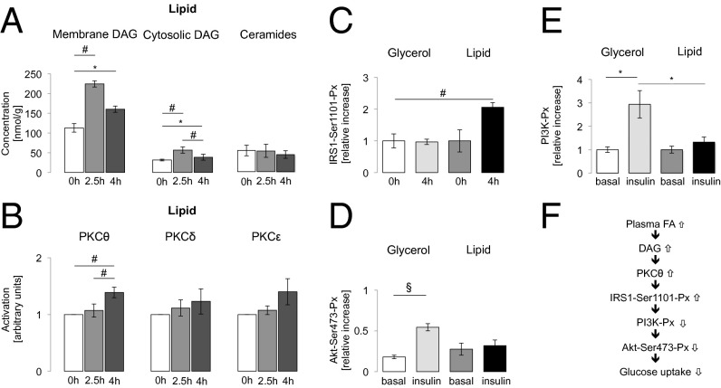
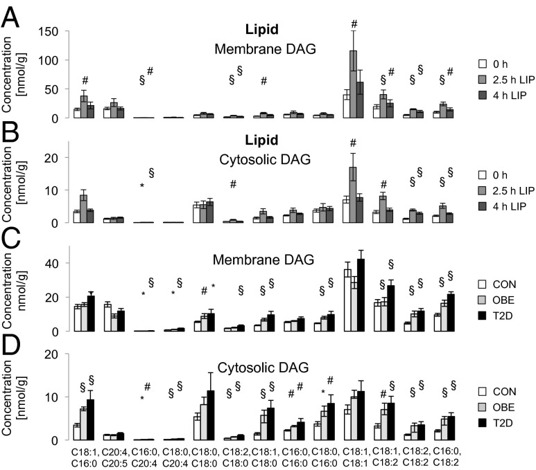
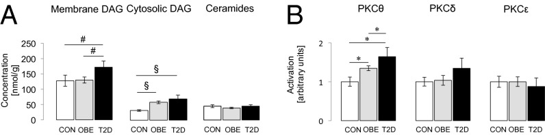
Muscle insulin resistance is a key feature of obesity and type 2 diabetes and is strongly associated with increased intramyocellular lipid content and inflammation. However, the cellular and molecular mechanisms responsible for causing muscle insulin resistance in humans are still unclear. To address this question, we performed serial muscle biopsies in healthy, lean subjects before and during a lipid infusion to induce acute muscle insulin resistance and assessed lipid and inflammatory parameters that have been previously implicated in causing muscle insulin resistance. We found that acute induction of muscle insulin resistance was associated with a transient increase in total and cytosolic diacylglycerol (DAG) content that was temporally associated with protein kinase (PKC)θ activation, increased insulin receptor substrate (IRS)-1 serine 1101 phosphorylation, and inhibition of insulin-stimulated IRS-1 tyrosine phosphorylation and AKT2 phosphorylation. In contrast, there were no associations between insulin resistance and alterations in muscle ceramide, acylcarnitine content, or adipocytokines (interleukin-6, adiponectin, retinol-binding protein 4) or soluble intercellular adhesion molecule-1. Similar associations between muscle DAG content, PKCθ activation, and muscle insulin resistance were observed in healthy insulin-resistant obese subjects and obese type 2 diabetic subjects. Taken together, these data support a key role for DAG activation of PKCθ in the pathogenesis of lipid-induced muscle insulin resistance in obese and type 2 diabetic individuals.
Keywords: insulin signaling; lipotoxicity.
Conflict of interest statement
The authors declare no conflict of interest.
Figures
References
-
- Cline GW, et al. Impaired glucose transport as a cause of decreased insulin-stimulated muscle glycogen synthesis in type 2 diabetes. N Engl J Med. 1999;341(4):240–246. - PubMed
-
- Roden M. How free fatty acids inhibit glucose utilization in human skeletal muscle. News Physiol Sci. 2004;19:92–96. - PubMed
Publication types
MeSH terms
Substances
Grants and funding
LinkOut - more resources
Full Text Sources
Other Literature Sources
Medical
Molecular Biology Databases
Miscellaneous
