

Designing a wearable navigation system for image-guided cancer resection surgery
- PMID: 24980159
- PMCID: PMC4332818
- DOI: 10.1007/s10439-014-1062-0
Designing a wearable navigation system for image-guided cancer resection surgery
Abstract
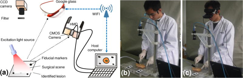
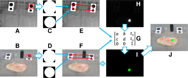
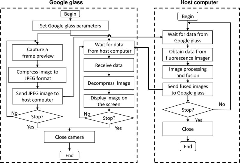
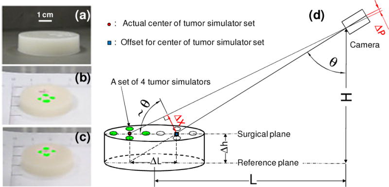
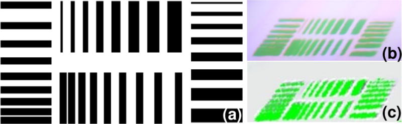
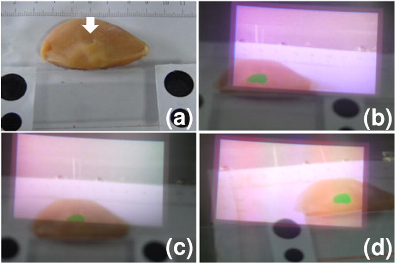
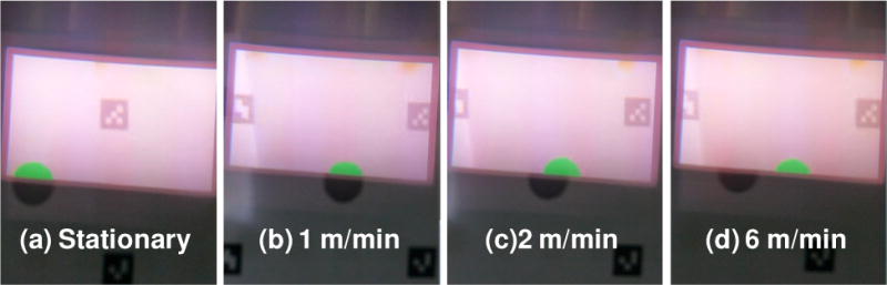
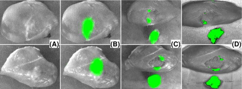
A wearable surgical navigation system is developed for intraoperative imaging of surgical margin in cancer resection surgery. The system consists of an excitation light source, a monochromatic CCD camera, a host computer, and a wearable headset unit in either of the following two modes: head-mounted display (HMD) and Google glass. In the HMD mode, a CMOS camera is installed on a personal cinema system to capture the surgical scene in real-time and transmit the image to the host computer through a USB port. In the Google glass mode, a wireless connection is established between the glass and the host computer for image acquisition and data transport tasks. A software program is written in Python to call OpenCV functions for image calibration, co-registration, fusion, and display with augmented reality. The imaging performance of the surgical navigation system is characterized in a tumor simulating phantom. Image-guided surgical resection is demonstrated in an ex vivo tissue model. Surgical margins identified by the wearable navigation system are co-incident with those acquired by a standard small animal imaging system, indicating the technical feasibility for intraoperative surgical margin detection. The proposed surgical navigation system combines the sensitivity and specificity of a fluorescence imaging system and the mobility of a wearable goggle. It can be potentially used by a surgeon to identify the residual tumor foci and reduce the risk of recurrent diseases without interfering with the regular resection procedure.
Figures
Comment in
-
Author's reply to the reader's comment on "designing a wearable navigation system for image-guided cancer resection surgery".Ann Biomed Eng. 2014 Dec;42(12):2602-3. doi: 10.1007/s10439-014-1157-7. Epub 2014 Oct 17. Ann Biomed Eng. 2014. PMID: 25323571 No abstract available.
-
Letter to the editor on "designing a wearable navigation system for image-guided cancer resection surgery".Ann Biomed Eng. 2014 Dec;42(12):2600-1. doi: 10.1007/s10439-014-1159-5. Epub 2014 Oct 17. Ann Biomed Eng. 2014. PMID: 25323572 No abstract available.
References
-
- Weber WP, Engelberger S, Viehl CT, Zanetti-Dallenbach R, Kuster S, et al. Accuracy of frozen section analysis versus specimen radiography during breast-conserving surgery for nonpalpable lesions. World Journal of Surgery. 2008;32:2599–606. - PubMed
-
- Waljee JF, Hu ES, Newman LA, Alderman AK. Predictors of re-excision among women undergoing breast-conserving surgery for cancer. Ann Surg Oncol. 2008;15:1297–303. - PubMed
-
- Horst KC, Smitt MC, Goffinet DR, Carlson RW. Predictors of local recurrence after breast-conservation therapy. Clin Breast Cancer. 2005;5:425–38. - PubMed
-
- Holland R, Veling SH, Mravunac M, Hendriks JH. Histologic multifocality of Tis, T1–2 breast carcinomas. Implications for clinical trials of breast-conserving surgery. Cancer. 1985;56:979–90. - PubMed
-
- Kuroiwa T, Kajimoto Y, Ohta T. Development of a fluorescein operative microscope for use during malignant glioma surgery: a technical note and preliminary report. Surg Neurol. 1998;50:41–8. discussion 8–9. - PubMed
Publication types
MeSH terms
Grants and funding
LinkOut - more resources
Full Text Sources
Other Literature Sources
Miscellaneous
