. 2014 Jul 1:14:60.
doi: 10.1186/1472-6750-14-60.

Optimized DNA extraction from neonatal dried blood spots: application in methylome profiling

Affiliations

Optimized DNA extraction from neonatal dried blood spots: application in methylome profiling

Akram Ghantous et al. BMC Biotechnol. .

Abstract

Background: Neonatal dried blood spots (DBS) represent an inexpensive method for long-term biobanking worldwide and are considered gold mines for research for several human diseases, including those of metabolic, infectious, genetic and epigenetic origin. However, the utility of DBS is restricted by the limited amount and quality of extractable biomolecules (including DNA), especially for genome wide profiling. Degradation of DNA in DBS often occurs during storage and extraction. Moreover, amplifying small quantities of DNA often leads to a bias in subsequent data, particularly in methylome profiles. Thus it is important to develop methodologies that maximize both the yield and quality of DNA from DBS for downstream analyses.

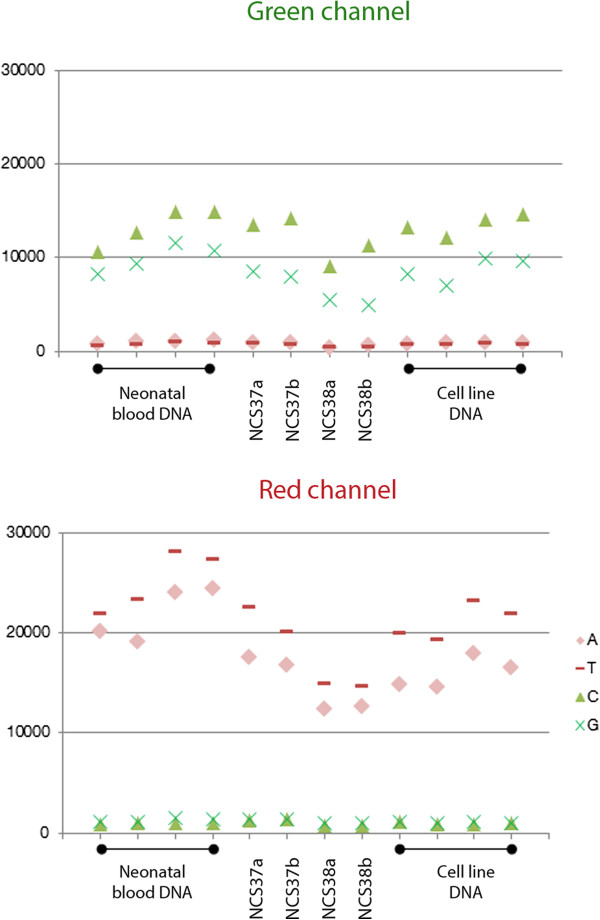
Results: Using combinations of in-house-derived and modified commercial extraction kits, we developed a robust and efficient protocol, compatible with methylome studies, many of which require stringent bisulfite conversion steps. Several parameters were tested in a step-wise manner, including blood extraction, cell lysis, protein digestion, and DNA precipitation, purification and elution. DNA quality was assessed based on spectrophotometric measurements, DNA detectability by PCR, and DNA integrity by gel electrophoresis and bioanalyzer analyses. Genome scale Infinium HumanMethylation450 and locus-specific pyrosequencing data generated using the refined DBS extraction protocol were of high quality, reproducible and consistent.

Conclusions: This study may prove useful to meet the increased demand for research on prenatal, particularly epigenetic, origins of human diseases and for newborn screening programs, all of which are often based on DNA extracted from DBS.


Figures

Figure 1
Figure 1
Phases and classification of protocols used to extract DNA from DBS. Two sequential phases, each encompassing three steps, are outlined (A) and were optimized in the different protocols or method combinations (B) used to extract DNA from DBS. A spin basket is shown next to Phase I.A and consists of a tube with an embedded perforated basket used to separate blood solutions from the filter papers from which they were extracted. A silica-gel column with a funnel-shape design is shown next to Phase II.E and often used to elute small volumes (5–30 μl), as manufactured by Macherey-Nagel and supplied with the extra-small (XS) versions of NucleoSpin kits (B).
Figure 2
Figure 2
DNA integrity and size range as assessed by agarose gel electrophoresis. (A) DNA size markers used to estimate size ranges are shown in addition to genomic DNA that was isolated from white blood cells (WBC) and used as a positive control. (B) Representative DBS from each of the tested protocols are shown, except for protocol QQ in which DNA amounts were insufficient to be analyzed by gel electrophoresis. Eight different gel sections are shown and are derived from either the same gel or different gels. In each section, two punches from the same NCS spot were run on the same gel, with the first punch, labeled ‘a’, representing protocol GQ and the second punch, labeled ‘b’, representing another unique protocol from the tested set. The two blue lines, representing the 100 and 1000 base pair (bp) size ranges, were set according to the molecular size marker used in each section. The 1000 base pair limit is a minimum size range with useful applications in many genetic and epigenetic studies, including Illumina’s HM450 Beadchip array. The results of other DBS analyzed by gel electrophoresis or bioanalyzer are summarized in Table 6.
Figure 3
Figure 3
HM450 QC plot using Non-polymorphic probes which assess overall performance. In the green channel, background signals are shown in red and pink while positive signals in opaque and fluorescent green. In the red channel, background signals are shown in opaque and fluorescent green while positive signals in red and pink. One non-polymorphic control has been designed for each of the four nucleotides A, T, C, and G. Four DBS DNA samples are shown between four neonatal blood and four cell line DNA samples, in each of the two plots. The DBS samples represent two NCS spots, 37 and 38, each consisting of two tested punches labeled ‘a’ or ‘b’.
Figure 4
Figure 4
Differential methylation and unsupervised clustering analysis of HM450 data from neonatal blood, DBS and cell line DNA. Neonatal blood and cell line DNA samples are used as positive controls of good DNA quality for reference comparisons with DNA extracted from DBS. Neonatal blood and DBS are from different individuals. Four DBS DNA samples are shown between four different neonatal blood and four different cell line DNA samples. The DBS samples represent two NCS spots, 37 and 38, each consisting of two tested punches labeled ‘a’ or ‘b’. HM450 beta-values were clustered using Euclidean distance as the dissimilarity index. As shown in the color key, the red and blue signals represent relatively hypomethylated and hypermethylated regions, respectively.
Figure 5
Figure 5
Methylation analyses of Line1 and AluYb8 loci using bisulfite pyrosequencing. The methylation levels of six and four CpG sites were analyzed for Line1(A) and AluYb8(B), respectively, and are expressed as percent of the total number of CpGs analyzed for each individual CpG site. The DBS samples represent two NCS spots, 37 and 38, each consisting of two tested punches labeled ‘a’ or ‘b’.

References

    1. Brown RC, Dwyer T, Kasten C, Krotoski D, Li Z, Linet MS, Olsen J, Scheidt P, Winn DM. Cohort profile: the International Childhood Cancer Cohort Consortium (I4C) Int J Epidemiol. 2007;36:724–730. - PubMed
    1. Guthrie R, Susi A. A simple phenylalanine method for detecting phenylketonuria in large populations of newborn infants. Pediatrics. 1963;32:338–343. - PubMed
    1. Mei JV, Alexander JR, Adam BW, Hannon WH. Use of filter paper for the collection and analysis of human whole blood specimens. J Nutr. 2001;131:1631S–1636S. - PubMed
    1. He H, Argiro L, Dessein H, Chevillard C. Improved technique that allows the performance of large-scale SNP genotyping on DNA immobilized by FTA technology. Infect Genet Evol. 2007;7:128–132. - PubMed
    1. Wong N, Morley R, Saffery R, Craig J. Archived Guthrie blood spots as a novel source for quantitative DNA methylation analysis. Biotechniques. 2008;45:423–424. 426, 428 passim. - PubMed

Publication types

LinkOut - more resources