. 2014 Jul 1:4:5519.
doi: 10.1038/srep05519.

An activator of mTOR inhibits oxLDL-induced autophagy and apoptosis in vascular endothelial cells and restricts atherosclerosis in apolipoprotein E⁻/⁻ mice

Affiliations

An activator of mTOR inhibits oxLDL-induced autophagy and apoptosis in vascular endothelial cells and restricts atherosclerosis in apolipoprotein E⁻/⁻ mice

Nan Peng et al. Sci Rep. .

Abstract

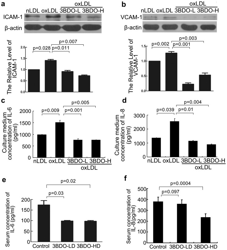
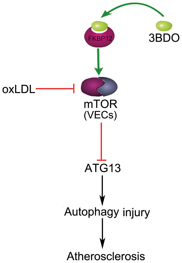
Oxidized low-density lipoprotein (oxLDL) inhibits mammalian target of rapamycin (mTOR) and induces autophagy and apoptosis in vascular endothelial cells (VECs) that play very critical roles for the cardiovascular homostasis. We recently defined 3-benzyl-5-((2-nitrophenoxy) methyl)-dihydrofuran-2(3H)-one (3BDO) as a new activator of mTOR. Therefore, we hypothesized that 3BDO had a protective role in VECs and thus stabilized atherosclerotic lesions in apolipoprotein E(-/-) (apoE(-/-)) mice. Our results showed that oxLDL inhibited the activity of mTOR and increased the protein level of autophagy-related 13 (ATG13) and its dephosphorylation, thus inducing autophagy in human umbilical vein endothelial cells (HUVECs). All of these effects were strongly inhibited by 3BDO. In vivo experiments confirmed that 3BDO activated mTOR and decreased the protein level of ATG13 in the plaque endothelium of apoE(-/-) mice. Importantly, 3BDO did not affect the activity of mTOR and autophagy in macrophage cell line RAW246.7 and vascular smooth muscle cells of apoE(-/-) mice, but suppressed plaque endothelial cell death and restricted atherosclerosis development in the mice. 3BDO protected VECs by activating mTOR and thus stabilized atherosclerotic lesions in apoE(-/-) mice.


Figures

Figure 1
Figure 1. Effect of 3BDO on mammalian target of rapamycin (mTOR) activity in oxidized low-density-lipoprotein (oxLDL)-treated human umbilical vein endothelial cells (HUVECs).
(a), Western blot analysis of mTOR, phosphorylated mTOR (p-mTOR, Ser2448), p70S6K, phosphorylated p70S6K (p-p70S6K, Thr389), 4EBP1, phosphorylated 4EBP1(p-4EBP1, Thr37/46) in HUVECs treated with native LDL (nLDL), 50 μg/ml; oxLDL, 50 μg/ml; 3BDO-L, 60 μM; and 3BDO-H, 120 μM, for 12 h (cropped, full-length blots are in Supplementary Figure S6). (b), Densitometry results of ratio of p-mTOR to total mTOR, p-p70S6K to total p70S6K and p-4EBP1 to total 4EBP1. nLDL control data were set to 1. Data are mean ± SEM, n = 3.
Figure 2
Figure 2. Effect of 3BDO on oxLDL-induced autophagy in HUVECs.
(a), Western blot analysis of ATG13 and p-ATG13 protein level and quantification (cropped, full-length blots are in Supplementary Figure S7), with nLDL, 50 μg/ml; oxLDL, 50 μg/ml; 3BDO-L, 60 μM; and 3BDO-H, 120 μM, for 12 h. nLDL control data were set to 1. Data are mean ± SEM; ATG13, *p = 0.017 vs. nLDL, #p = 0.024 vs. oxLDL, & p = 0.024 vs. oxLDL; p-ATG13/ATG13, **p = 0.004 vs. nLDL, #p = 0.039 vs. oxLDL, & p = 0.011vs. oxLDL. n = 3. (b), Western blot analysis of LC3 –II and p62 and quantification (cropped, full-length blots are in Supplementary Figure S8), with nLDL, 50 μg/ml; oxLDL, 50 μg/ml; 3BDO-L, 60 μM; and 3BDO-H, 120 μM, for 12 h. Protein levels were normalized to that of β-actin. nLDL control data were set to 1. Data are mean ± SEM; LC3–II, **p = 0.0008 vs. nLDL, #p = 0.01 vs. oxLDL, &&p = 0.0005 vs. oxLDL; p62, **p = 0.004 vs. nLDL, ##p = 0.003 vs. oxLDL, &&p = 0.009 vs. oxLDL. n = 3. (c), Immunofluorescence staining of LC3, with nLDL, 50 μg/ml; oxLDL, 50 μg/ml; 3BDO-L, 60 μM, for 12 h. Bar = 5 μM. Bar charts showed the quantification of average endogenous LC3 puncta per cell. Different fields of view (>3 regions) were analyzed on the microscope for each labeling condition, and representative results are shown. Data are mean ± SEM.
Figure 3
Figure 3. 3BDO activated mTOR in the endothelium of apoE-/- mice.
(a), Double-stained images of co-localization (yellow) of p-p70S6K with CD31-positive VECs of apoE-/- mice with and without 3BDO treatment. Bar = 60 μM. (b), Quantification of p-p70S6K protein level in a. Data are mean ± SEM. n = 6.
Figure 4
Figure 4. 3BDO increased the level of ATG13 in the endothelium of apoE-/- mice.
(a), Double-stained images of co-localization (yellow) of ATG13 with CD31-positive VECs of apoE-/- mice with and without 3BDO treatment. Bar = 60 μM. (b), Quantification of ATG13 protein level in a. Data are mean ± SEM. n = 6.
Figure 5
Figure 5. 3BDO inhibited endothelium autophagy and apoptosis in apoE-/- mice.
(a), En face staining of LC3 patches (green) in the thoracic aorta endothelium of apoE-/- mice. Endothelial cells were marked with CD31 (red). Nucleus (blue) was stained with DAPI. Bar = 60 μM. (b), Quantification of LC3 dots in the thoracic aorta endothelium. Data are mean ± SEM. n = 6. (c), Sections from the mice aortic roots were labeled by TUNEL to detect apoptotic cells and counterstained with DAPI to detect nuclei. The arrows indicated the TUNEL-positive cells. Bar = 60 μM. (d), Quantification of TUNEL-positive cells in the plaque endothelium. Data are mean ± SEM. n = 6.
Figure 6
Figure 6. Effect of 3BDO on the phenotype of aortic atherosclerotic plaque in apoE-/- mice.
(a), Oil-red O staining of whole aortas and quantification. Data are mean ± SEM, Bar = 3 mm, n = 6. (b), Hematoxylin and eosin (H&E) staining and quantification of plaque area. Data are mean ± SEM. Bar = 500 μm. (c), From the top to bottom panel, Oil-red O staining of atherosclerotic lesions, immunofluorescence staining for mouse α-smooth muscle actin, Mac-3 (M3/84) and in situ zymography of matrix metalloproteinase 2/9 (MMP-2/9) activity. Bar for Oil-red O staining = 500 μm, others = 80 μm. D, Quantification of lipid area, smooth muscle area, macrophage area, and MMP-2/9 activity in control and 3BDO-treated groups. Data are mean ± SEM. n = 6.
Figure 7
Figure 7. Effect of 3BDO on inflammatory response in oxLDL-treated HUVECs and apoE-/- mice.
(a) and (b), Western blot analysis of the effect of 3BDO on intracellular adhesion molecule 1 (ICAM-1) and vascular cell adhesion molecule 1 (VCAM-1) level (full-length blots are in Supplementary Figure S9 and S10), with oxLDL, 50 μg/ml; 3BDO-L, 60 μM; 3BDO-H, 120 μM for 12 h. Protein levels were normalized to that of β-actin. Data are mean ± SEM. n = 3. (c) and (d), ELISA of the secretion of interleukin 6 (IL-6) and IL-8 from HUVECs with 3BDO treatment. oxLDL, 50 μg/ml; 3BDO-L, 60 μM; 3BDO-H, 120 μM for 12 h. Data are mean ± SEM. n = 3. (e) and (f), ELISA of the secretion of interleukin 6 (IL-6) and IL-8 in serum of apoE-/- mice. Data are mean ± SEM. n = 6.
Figure 8
Figure 8. Conceptual schematic of 3BDO and mTOR action mechanism in atherosclerosis.
OxLDL could induce mTOR inhibition, and the inhibition of mTOR contributes to autophagy by increasing the protein level of ATG13 and inhibiting ATG13 phosphorylation. The excessively stimulated autophagy finally leads to endothelial injury of apoE-/- mice. 3BDO could reverse oxLDL-induced mTOR inhibition in VECs and inhibit oxLDL-induced ATG13 increase and dephosphorylation, thus relieving oxLDL-induced autophagy injury and inhibiting atherosclerosis development in apoE-/- mice.

References

    1. Rioufol G. et al. Multiple atherosclerotic plaque rupture in acute coronary syndrome: a three-vessel intravascular ultrasound study. Circulation 106, 804–808 (2002). - PubMed
    1. Simionescu M. & Antohe F. Functional ultrastructure of the vascular endothelium: changes in various pathologies. Handb. Exp. Pharmacol. 176 Pt 1, 41–69 (2006). - PubMed
    1. Sima A. V., Stancu C. S. & Simionescu M. Vascular endothelium in atherosclerosis. Cell. Tissue. Res. 335, 191–203 (2009). - PubMed
    1. Rajendran P. et al. The Vascular Endothelium and Human Diseases. Int. J. Biol. Sci. 9, 1057–1069 (2013). - PMC - PubMed
    1. Weichhart T. Mammalian target of rapamycin: a signaling kinase for every aspect of cellular life. Methods. Mol. Biol. 821, 1–14 (2012). - PubMed

Publication types

MeSH terms

Substances