. 2014 Aug 5;22(8):1090-1104.
doi: 10.1016/j.str.2014.05.009. Epub 2014 Jun 26.

Structure of the C. elegans ZYG-1 cryptic polo box suggests a conserved mechanism for centriolar docking of Plk4 kinases

Affiliations

Structure of the C. elegans ZYG-1 cryptic polo box suggests a conserved mechanism for centriolar docking of Plk4 kinases

Ekaterina Shimanovskaya et al. Structure. .

Abstract

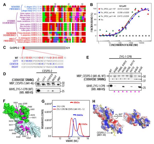
Plk4 family kinases control centriole assembly. Plk4s target mother centrioles through an interaction between their cryptic polo box (CPB) and acidic regions in the centriolar receptors SPD-2/Cep192 and/or Asterless/Cep152. Here, we report a crystal structure for the CPB of C. elegans ZYG-1, which forms a Z-shaped dimer containing an intermolecular β sheet with an extended basic surface patch. Biochemical and in vivo analysis revealed that electrostatic interactions dock the ZYG-1 CPB basic patch onto the SPD-2-derived acidic region to promote ZYG-1 targeting and new centriole assembly. Analysis of a different crystal form of the Drosophila Plk4 (DmPlk4) CPB suggests that it also forms a Z-shaped dimer. Comparison of the ZYG-1 and DmPlk4 CPBs revealed structural changes in the ZYG-1 CPB that confer selectivity for binding SPD-2 over Asterless-derived acidic regions. Overall, our findings suggest a conserved mechanism for centriolar docking of Plk4 homologs that initiate daughter centriole assembly.


Figures

Figure 1
Figure 1. The ZYG-1 CPB forms a Z-shaped end-to-end dimer with a conserved basic patch
(A) Ribbon diagram of the ZYG-1 CPB color-ramped from blue to red (N- to C-terminus). The two polo boxes (PB1 and PB2) are indicated and the two disordered loops in PB2 are shown as dashed lines. (B) Crystal-packing suggests two possible dimer interfaces. (C) SAXS of the ZYG-1 CPB in solution. The logarithm of the scattering intensity is plotted as a function of momentum transfer s = 4π sin(θ)/λ, where θ is the scattering angle and λ is the X-ray wavelength. Inset shows the boxed region at higher magnification. (D) Two views of the representative ab initio low resolution shape reconstructed by GASBOR. The most probable model, chosen from a dozen reconstructions by DAMAVER (Volkov & Svergun, 2003), is shown (transparent beads) superimposed with the Z-dimer structure (green ribbons). The overall normalized spatial discrepancy (NSD) was 1.3 and all models were consistent. (E-H) Surface plots of the ZYG-1 CPB Z-dimer highlighting conserved basic (blue) and acidic (magenta) residues (E, G) and electrostatic potential (F, H). Views showing the basic patch (E, F; dashed line in F) and the reverse side (G, H) of the dimer are shown.
Figure 2
Figure 2. The ZYG-1 CPB interacts with the CeSPD-2 N-terminus through a series of electrostatic interactions
(A) Acidic regions in SPD-2 and asterless homologs were manually selected and aligned using Muscle WS in Jalview. Color code: negatively charged (red), positively charged (blue), aromatic (gold), hydrophilic (green), conformationally special (pink) and cysteine (yellow). (B) MST binding curves of thioredoxin (Trx) fusions with regions of the CeSPD-2 N-terminus to the ZYG-1 CPB. (C) Schematic showing the CeSPD-2 AR (red box) and residue changes in charge reversal mutants. (D, E) In vitro pull-down experiments. Resin-immobilized WT or mutant MBP-CeSPD-2 AR (aa 1-46) (coomassie gels; top panels) were used to pull down WT or mutant 6×His-ZYG-1 CPB (Western blots; bottom panels). Asterisks mark MBP released due to degradation. Equivalent solubility of all ZYG-1 CPB mutants in (E) was independently confirmed. Magenta arrowheads mark CPB mutants that reduced the interaction. (F) Location of positively charged residues on the ZYG-1 CPB dimer that contribute to the interaction with the CeSPD-2 AR. (G) Static light scattering results for the ZYG-1 CPB alone (blue) or mixed in a 1:2 ratio with Trx fusion with the CeSPD-2 N-terminus. (H) In silico docking result using the ZYG-1 CPB dimer (electrostatic surface plot) as receptor and the CeSPD-2 N-terminus (aa 11-44; yellow stick model) as ligand.
Figure 3
Figure 3. A distributed array of negatively charged residues in the CeSPD-2 N-terminus recruits ZYG-1 to mother centrioles to initiate centriole duplication in vivo
(A) Single copy mCherry::spd-2RR transgene. (B) Western blot of mCherry::spd-2RR worm lysates, with (+) or without (−) endogenous SPD-2 depletion, probed for SPD-2 (top) and α-tubulin (bottom). Asterisk marks a background band. (C) (left) Confocal images of one-cell stage embryos expressing mCherry::SPD-2 fusions. (right) Graph of mean centrosomal mCherry::SPD-2 fluorescence for the indicated strains (n = number of centrosomes; error bars = SEM). (D, E) Deconvolved immunofluorescence images of first (left) and second division (right) embryos lacking (D) or expressing the indicated spd-2 transgenes (E). Embryos were depleted of the indicated protein (D) or endogenous SPD-2 (E) by RNAi and were stained for microtubules (green) and DNA (red). n = number of embryos imaged. (F) Plots showing percent embryonic viability (left) and frequency of second division monopolar spindles (right). Error bars = SD of percent lethality per worm; N = number of worms, n = total number of embryos (left) or second division cells (right). (G) Deconvolved immunofluorescence images of embryos stained for DNA and SAS-4 (red and green; left panels) and ZYG-1 (right panels). Embryos expressing the indicated spd-2 transgenes were depleted of endogenous SPD-2 by RNAi. (H) Graph plotting mean centrosomal ZYG-1 fluorescence. Values are percentage of the mean control value. n = number of centrosomes. Error bars = SEM. Scale bars, 10 m.
Figure 4
Figure 4. Basic patch residues on the ZYG-1 CPB dimer are required to target ZYG-1 to mother centrioles and promote daughter centriole assembly
(A) Single copy zyg-1RR transgene and predicted ZYG-1 protein carrying the temperature sensitive P442L mutation. (B) Surface plot of the ZYG-1 CPB dimer showing the three tested mutant clusters. (C) Western blot of lysates from worms lacking a transgene (No Transgene) or with the indicated zyg-1RR transgenes with (+) or without (−) endogenous ZYG-1 depletion. The blot was probed for ZYG-1 (top) and α-tubulin (bottom). Asterisks mark variable background bands due to the bacterial food that the worms eat. Serial dilutions of lysate prepared from worms without a transgene (first five lanes) were used to assess endogenous ZYG-1 depletion. Numbers are percentage loaded relative to the 100% no transgene control. (D, E) Plots showing percent embryonic viability (D) and the frequency of second division monopolar spindles (E). Error bars in D = SD of percent lethality per worm; N = number of worms, n = total number of embryos (D) or the number second division cells (E). (F) (left) Deconvolved immunofluorescence images of control and zyg-1(RNAi) embryos stained for DNA and SAS-4 (red and green; left panels) and ZYG-1 (right panels). (right) Graph plotting mean centrosomal ZYG-1 fluorescence. Values are percentage of the mean control value. n = number of centrosomes. Error bars = SEM. (G) (left) Representative deconvolved widefield immunofluorescence images of embryos stained for DNA and SAS-4 (red and green; left panels) and ZYG-1 (right panels). Embryos expressing the indicated zyg-1 transgenes were depleted of endogenous ZYG-1 by RNAi. (right) Graph plotting mean centrosomal ZYG-1 fluorescence. Values are percentage of the mean control value. n = number of centrosomes. Error bars = SEM. Scale bars, 10 m.
Figure 5
Figure 5. The DmPlk4 CPB forms a Z-shaped end-to-end dimer analogous to that formed by the ZYG-1 CPB
(A) Ribbon diagram of the DmPlk4 CPB structure, color-ramped from blue to red (N- to C-terminus). The disordered loop (aa 549-552) in PB2 is shown as a dashed line. (B, C) Top (B) and side (C) views of a single hexagonal unit in the DmPlk4 CPB crystal, which contains 12 CPBs forming 6 pairs (yellow, blue, pink, green, red, and cyan) arranged in a spiral along the vertical screw axis (c-axis). (D) Side view of the previously reported butterfly-like side-by-side dimer (Slevin et al., 2012). (E) The inter-molecular contact mediated by the β5-β6 loops (arrows) of adjacent PB1 domains in the hexagonal unit. (F) The Z-dimer formed by the DmPlk4 CPB. (G, H) Superposition of the Z-dimer (G) and butterfly-dimer (H) onto the ab-initio shell reconstructed from SAXS data. The ab inito model is the most representative model selected by DAMAVER out of 40 DAMMIN runs. The mean NSD was 0.542. (I) SAXS analysis of the DmPlk4 CPB in solution. Inset shows the boxed region at a higher magnification.
Figure 6
Figure 6. The SPD-2/Cep192 acidic region interacts with a more compact region of the MmPlk4 CPB than the asterless/Cep152 acidic region
(A) Surface plot (front view) of the DmPlk4 CPB Z-dimer with the two CPBs shown in green and pale cyan. Completely and highly (80-95% identity) conserved basic (dark and light blue) and acidic (red and pink) residues are shown. Residues in the basic surface patch (dashed line) are labeled. (B) In silico docking result using the DmPlk4 CPB Z-dimer (electrostatic surface plot) as receptor and the DmAsl N-terminus (aa 21-60; yellow stick model) as ligand. (C) Location of positively charged residues on the ZYG-1 CPB dimer that contribute to the interaction with the CeSPD-2 AR, repeated from Figure 2F for comparison. An end view is shown on right. (D-F) In vitro pull down experiments. (D, left) Resin-immobilized WT or mutant 6×His-DmPlk4 CPB (Coomassie gel; top panel) was used to pull down MBP-DmAsl AR (aa 21-60; Western blot; bottom panel). (E-F, left) Resin-immobilized GST-MmAsl/Cep152 AR (E) or GST-MmSPD-2/Cep192 AR (F) (Coomassie gels; top panels) were used to pull down WT and mutant versions of the MBP-MmPlk4 CPB (Western blots; bottom panels). (D-F, right) Location of positively charged residues on the DmPlk4 CPB dimer (D) or the modeled MmPlk4 CPB dimer (E,F; see Figure S5H) whose mutation reduced (magenta) or did not affect (pale yellow) interaction with the tested ARs. Arrows indicate a mutant outside the conserved patch that did not significantly affect the binary interactions. Empty triangles mark mutants that significantly enhanced interactions.
Figure 7
Figure 7. Structural changes in the ZYG-1 CPB dimer confer the ability to selectively bind SPD-2-derived acidic regions
(A) Kds measured by MST for different CPB-AR pairs. (B) Superposition of the DmPlk4 and ZYG-1 CPB dimers aligning one of the two CPBs. (left) A side view demonstrates that the ZYG-1 CPB dimer bends 35° away from the long axis relative to the DmPlk4 dimer. (right) A view of the plane containing the intermolecular β-sheet shows that the second ZYG-1 CPB is rotated counterclockwise towards the long axis by 15°. (C,D) (left) Electrostatic surface plots showing side and top views of the DmPlk4 (C) and ZYG-1 (D) CPB dimers. The DmPlk4 dimer has a linear long axis and the blunt angle at the dimeric junction is 105°. The ZYG-1 CPB dimer is curved, with a 35° distortion of the long axis and a blunt angle of 120°. (right) The distributions of basic residues involved in the DmAsl AR-DmPlk4 and CeSPD-2 AR-ZYG-1 interactions are shown for comparison. (E) Schematics illustrating docking modes for asterless and SPD-2-derived acidic regions onto the ZYG-1 and Plk4 CPB dimers. The two CPBs in each dimer are colored green and pale cyan. Conserved basic clusters are shown as blue patches. Acidic regions are shown as curved lines with the rough positions of the acidic (red) and basic (blue) segments highlighted. (F) Sequence comparison of the acidic regions in asterless and SPD-2 homologs. Acidic (red) and basic (blue) residues are highlighted.

Comment in

  • Polo-like kinase 4 shapes up.
    Levine MS, Holland AJ. Levine MS, et al. Structure. 2014 Aug 5;22(8):1071-1073. doi: 10.1016/j.str.2014.07.004. Structure. 2014. PMID: 25099950

References

    1. Anderhub SJ, Krämer A, Maier B. Centrosome amplification in tumorigenesis. Cancer Lett. 2012;322:8–17. doi:10.1016/j.canlet.2012.02.006. - PubMed
    1. Azimzadeh J, Marshall WF. Building the centriole. Curr. Biol. 2010;20:R816–25. doi:10.1016/j.cub.2010.08.010. - PMC - PubMed
    1. Brito DA, Gouveia SM, Bettencourt-Dias M. Deconstructing the centriole: structure and number control. Curr. Opin. Cell Biol. 2012;24:4–13. doi:10.1016/j.ceb.2012.01.003. - PubMed
    1. Carvalho-Santos Z, Azimzadeh J, Pereira-Leal JB, Bettencourt-Dias M. Evolution: Tracing the origins of centrioles, cilia, and flagella. J. Cell Biol. 2011;194:165–175. doi:10.1083/jcb.201011152. - PMC - PubMed
    1. Chen C-C, Hwang J-K, Yang J-M. PS)2-v2: template-based protein structure prediction server. BMC Bioinformatics. 2009;10:366. doi:10.1186/1471-2105-10-366. - PMC - PubMed

Publication types

MeSH terms

Substances

LinkOut - more resources