. 2014 Sep;65(17):5033-47.
doi: 10.1093/jxb/eru266. Epub 2014 Jun 30.

Experimental and bioinformatic characterization of a recombinant polygalacturonase-inhibitor protein from pearl millet and its interaction with fungal polygalacturonases

Affiliations

Experimental and bioinformatic characterization of a recombinant polygalacturonase-inhibitor protein from pearl millet and its interaction with fungal polygalacturonases

S Ashok Prabhu et al. J Exp Bot. 2014 Sep.

Abstract

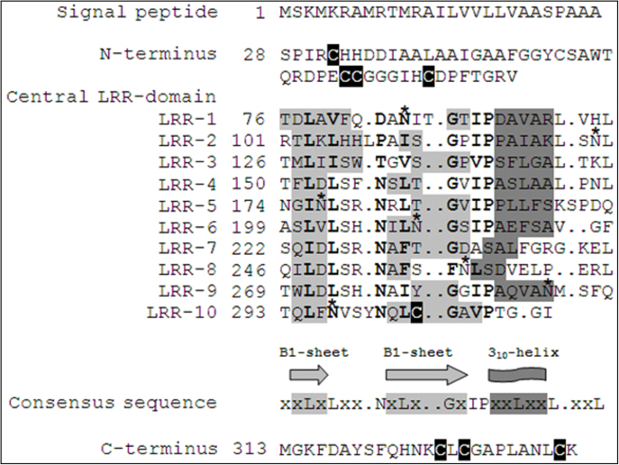
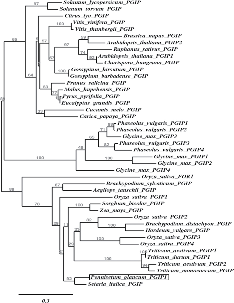
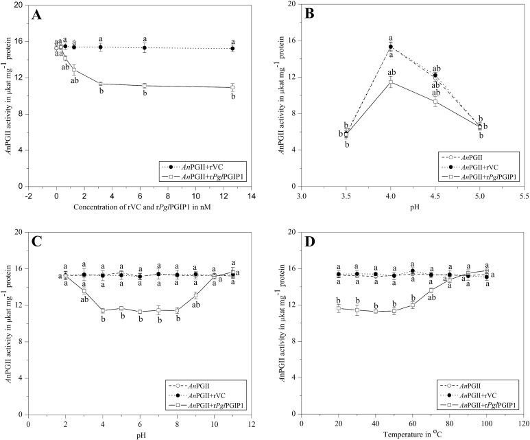
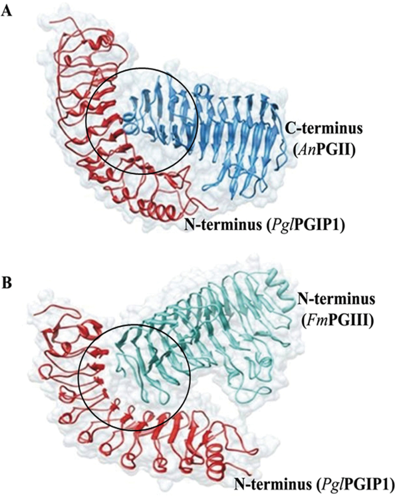
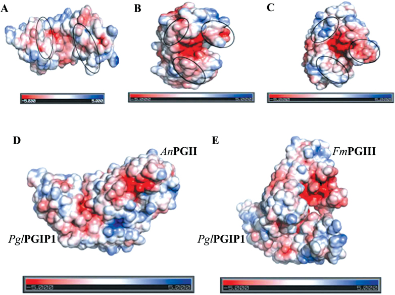
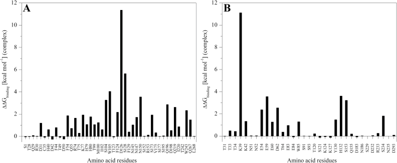
Polygalacturonases (PGs) are hydrolytic enzymes employed by several phytopathogens to weaken the plant cell wall by degrading homopolygalacturonan, a major constituent of pectin. Plants fight back by employing polygalacturonase-inhibitor proteins (PGIPs). The present study compared the inhibition potential of pearl millet PGIP (Pennisetum glaucum; PglPGIP1) with the known inhibition of Phaseolus vulgaris PGIP (PvPGIP2) against two PGs, the PG-II isoform from Aspergillus niger (AnPGII) and the PG-III isoform from Fusarium moniliforme (FmPGIII). The key rationale was to elucidate the relationship between the extent of sequence similarity of the PGIPs and the corresponding PG inhibition potential. First, a pearl millet pgip gene (Pglpgip1) was isolated and phylogenetically placed among monocot PGIPs alongside foxtail millet (Setaria italica). Upstream sequence analysis of Pglpgip1 identified important cis-elements responsive to light, plant stress hormones, and anoxic stress. PglPGIP1, heterologously produced in Escherichia coli, partially inhibited AnPGII non-competitively with a pH optimum between 4.0 and 4.5, and showed no inhibition against FmPGIII. Docking analysis showed that the concave surface of PglPGIP1 interacted strongly with the N-terminal region of AnPGII away from the active site, whereas it weakly interacted with the C-terminus of FmPGIII. Interestingly, PglPGIP1 and PvPGIP2 employed similar motif regions with few identical amino acids for interaction with AnPGII at non-substrate-binding sites; however, they engaged different regions of AnPGII. Computational mutagenesis predicted D126 (PglPGIP1)-K39 (AnPGII) to be the most significant binding contact in the PglPGIP1-AnPGII complex. Such protein-protein interaction studies are crucial in the future generation of designer host proteins for improved resistance against ever-evolving pathogen virulence factors.

Keywords: Computational mutagenesis; PGIPs; PGs; Phaseolus vulgaris; electrostatic surface potential; inhibition studies; pearl millet; protein modelling and docking..


Figures

Fig. 1.
Fig. 1.
Sequence organization of derived amino acid sequences of PglPGIP1. The displayed sequence organization is a result of alignment of PglPGIP1 with PvPGIP2 whose secondary structure has been determined (Di Matteo et al., 2003). The LRR consensus sequence ‘xxLxLxx.NxLx..GxIPxxLxxL.xxL’ is shown. Putative residues contributing to form the secondary structure elements are based on PvPGIP2 and indicated in light grey (sheets B1 and B2) and dark grey (310-helix). Putative glycosylation sites are indicated by an asterisk. The conserved C residues are highlighted in black. Numbering of amino acid residues is shown on the left.
Fig. 2.
Fig. 2.
Phylogenetic tree showing the affiliation of PglPGIP1 among other known monocot and dicot PGIPs. The deduced amino acid sequences of PglPGIP1 and other known PGIP sequences obtained from GenBank were aligned using MUSCLE version 3.7, curated using the Gblocks version 0.91b, and then submitted to PhyML version 3.0 aLRT for phylogenetic analysis, and the tree was rendered using TreeDyn version 198.3. The position of PglPGIP1 is highlighted by a box. The branch support values are represented at branch points and the branch length scale is shown below the tree. The protein accession numbers are summarized in Supplementary Table S1.
Fig. 3.
Fig. 3.
AnPGII inhibition assay. (A) Effect of inhibitor concentration. AnPGII (5ng) was assayed with and without inhibitor (rPglPGIP1/rVC) over a concentration range of 0.316–12.64nM and a graph with the enzyme activity over inhibitor concentration was plotted. (B) pH optimum. AnPGII (5ng) was assayed with and without inhibitor at a concentration (rPglPGIP1/rVC) of 3.16nM and a graph with the enzyme activity over pH units was plotted to determine the pH optima of inhibition. (C) pH stability. AnPGII (5ng) was assayed with and without inhibitor pre-incubated for 16h at pH values of 2.0–10.0 at 4 °C upon reconstitution in the assay buffer [at a concentration (rPglPGIP1/rVC) of 3.16 nM] and a graph with the enzyme activity over pH units was plotted to determine the pH stability of inhibitor. (D) Temperature stability. AnPGII (5ng) was assayed with and without inhibitor pre-incubated for 1h at temperatures ranging from 20 to 100 °C [at a concentration (rPglPGIP1/rVC) of 3.16 nM] and a graph with the enzyme activity over temperature was plotted to determine the temperature stability of inhibitor. The data points are means of a single experiment carried out in triplicates. Results are shown as means±standard error. Means designated with the same letter are not significantly different according to Tukey’s HSD test at P<0.05.
Fig. 4.
Fig. 4.
Protein docking analysis. Docked poses of PglPGIP1–AnPGII (A) and PglPGIP1–FmPGIII (B) complexes. PglPGIP1 interacts through its solvent-exposed concave cavity with AnPGII and FmPGIII at their N- and C-termini (circled in black), respectively. The substrate-binding site in FmPGIII appears to be more exposed compared with that of AnPGII. (This figure is available in colour at JXB online.)
Fig. 5.
Fig. 5.
Electrostatic surface potential of individual proteins and protein complexes. Electrostatic potential maps of PglPGIP1 (A), AnPGII (B), FmPGIII (C), PglPGIP1–AnPGII (D), and PglPGIP1–FmPGIII (E) complexes on which surface colours are fixed at red (–5) or blue (+5). Marked regions display the difference in charge distributions in surface maps of the individual proteins.
Fig. 6.
Fig. 6.
Computational alanine mutagenesis of PglPGIP1–AnPGII interface residues. The plot displays the contribution of individual interacting residues from PglPGIP1 (the PGIP residue numbering followed excludes the putative signal peptide) (A) and AnPGII (B) in the stability of the PglPGIP1–AnPGII complex. Interface residues were defined as those residues with a side chain having at least one atom within a sphere with 4 Å radius of an atom belonging to the other partner in the complex and binding hotspots defined as those residues that show ∆∆Gbinding >1 kcal mol–1.

Similar articles

Cited by

References

    1. Abu-Goukh AA, Greve LC, Labavitch JM. 1983. Purification and partial characterization of “Barlett” pear fruit polygalacturonase inhibitors. Physiological Plant Pathology 23, 111–122
    1. Abu-Goukh AA, Labavitch JM. 1983. The in vivo role of “Bartlett” pear fruit polygalacturonase inhibitors. Physiological Plant Pathology 23, 123–135
    1. Ahsan N, Yoon HS, Jo J. 2005. Molecular cloning of a BcPGIP cDNA from Brassica campestris and its expression to several stresses. Plant Science 169, 1081–1089
    1. Anisimova M, Gascuel O. 2006. Approximate likelihood-ratio test for branches: a fast, accurate, and powerful alternative. Systematic Biology 55, 539–552 - PubMed
    1. Anthon GE, Barrett DM. 2002. Determination of reducing sugars with 3-methyl-2-benzothiazolinonehydrazone. Analytical Biochemistry 305, 287–289 - PubMed

Publication types