. 2014 Sep;82(9):3867-79.
doi: 10.1128/IAI.01769-14. Epub 2014 Jun 30.

Two functional type VI secretion systems in avian pathogenic Escherichia coli are involved in different pathogenic pathways

Affiliations

Two functional type VI secretion systems in avian pathogenic Escherichia coli are involved in different pathogenic pathways

Jiale Ma et al. Infect Immun. 2014 Sep.

Erratum in

Abstract

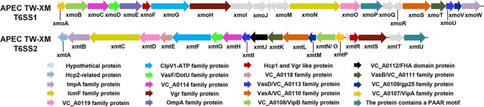
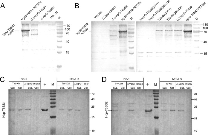
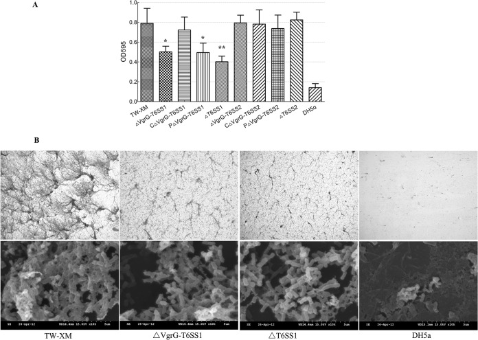
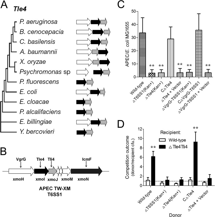
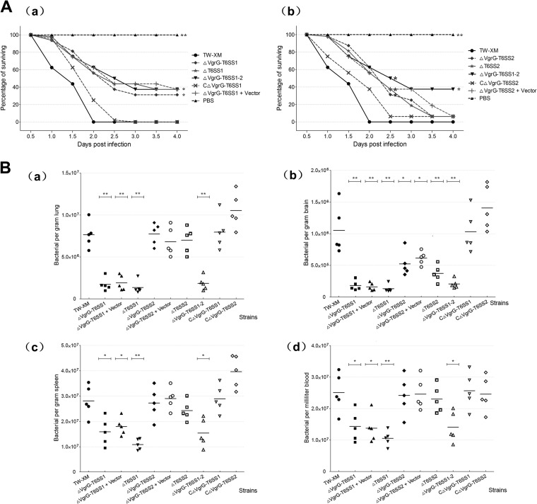
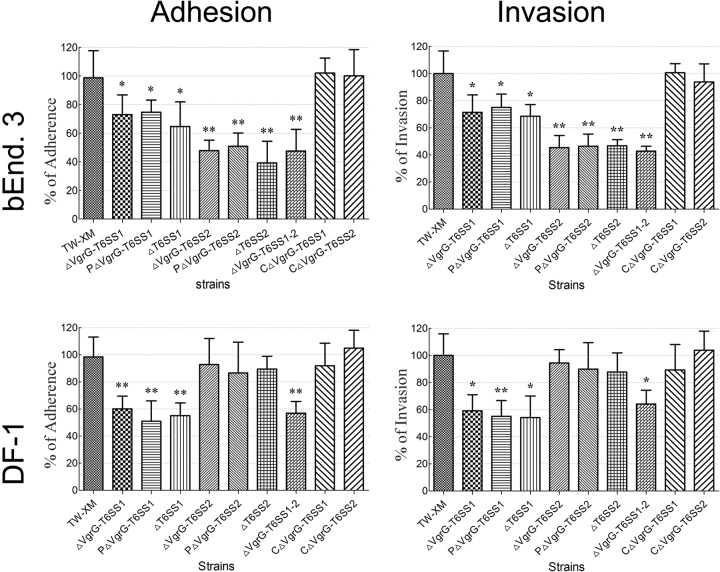
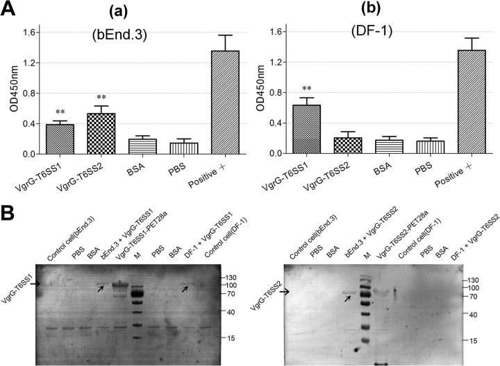
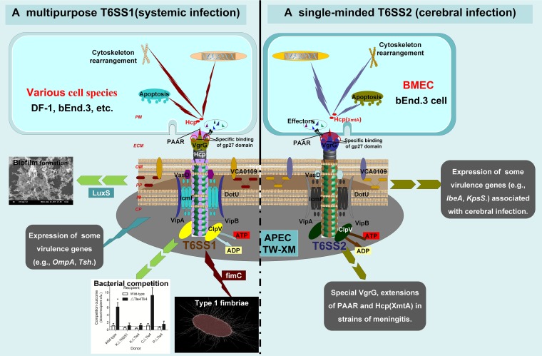
Type VI secretion systems (T6SSs) are involved in the pathogenicity of several Gram-negative bacteria. The VgrG protein, a core component and effector of T6SS, has been demonstrated to perform diverse functions. The N-terminal domain of VgrG protein is a homologue of tail fiber protein gp27 of phage T4, which performs a receptor binding function and determines the host specificity. Based on sequence analysis, we found that two putative T6SS loci exist in the genome of the avian pathogenic Escherichia coli (APEC) strain TW-XM. To assess the contribution of these two T6SSs to TW-XM pathogenesis, the crucial clpV clusters of these two T6SS loci and their vgrG genes were deleted to generate a series of mutants. Consequently, T6SS1-associated mutants presented diminished adherence to and invasion of several host cell lines cultured in vitro, decreased pathogenicity in duck and mouse infection models in vivo, and decreased biofilm formation and bacterial competitive advantage. In contrast, T6SS2-associated mutants presented a significant decrease only in the adherence to and invasion of mouse brain microvascular endothelial cell (BMEC) line bEnd.3 and brain tissue of the duck infection model. These results suggested that T6SS1 was involved in the proliferation of APEC in systemic infection, whereas VgrG-T6SS2 was responsible only for cerebral infection. Further study demonstrated that VgrG-T6SS2 was able to bind to the surface of bEnd.3 cells, whereas it did not bind to DF-1 (chicken embryo fibroblast) cells, which further proved the interaction of VgrG-T6SS2 with the surface of BMECs.


Figures

FIG 1
FIG 1
Schematic diagram of the genetic organization of TW-XM T6SS gene clusters. Genes encoding conserved domain proteins are represented by the same colors. Silver arrows refer to other genes included in the T6SS loci that were not identified as part of the conserved core described by Boyer et al. (57). The direction of the arrows indicates the direction of transcription. The color keys for the functional classes of genes in the T6SS loci are shown at the bottom. The database of Clusters of Orthologous Groups of proteins was obtained from the NCBI.
FIG 2
FIG 2
Expression levels of VgrGs and Hcps in different strains were studied by immunoblotting. The quality inspection of protein samples was demonstrated in Fig. S2 in the supplemental material. (A) VgrG-T6SS1 was not detected in the lysate from mutant strain ΔVgrG-T6SS1. The expression of vgrG-T6SS1 was demonstrated by Western blotting with VgrG-T6SS1 antibody using the lysates of wild-type, ΔVgrG-T6SS1, and CΔVgrG-T6SS1 strains cultured in DMEM. The recombinant VgrG-T6SS1 was used as a positive control. (B) The expression of vgrG-T6SS2 must be activated by host cells. Data are from Western blotting with VgrG-T6SS2 antibody using the lysates of wild-type, ΔVgrG-T6SS2, and CΔVgrG-T6SS2 strains cultured in DMEM and coincubated with DF-1 or bEnd.3 cells. The recombinant VgrG-T6SS2 was used as a positive control. (C and D) Lanes Sup., supernatants from wild-type, ΔVgrG-T6SS1, and ΔVgrG-T6SS2 strains coincubated with DF-1 or bEnd.3 cells; lanes Cell, cytosol of DF-1 or bEnd.3 cells coincubated with wild-type, ΔVgrG-T6SS1, and ΔVgrG-T6SS2 strains. (C) Hcp-T6SS1 is undetected in cytosol of DF-1 and bEnd.3 cells coincubated with mutant strain ΔVgrG-T6SS1. The result was demonstrated by Western blotting with Hcp-T6SS1 antibody. (D) Hcp-T6SS2 is undetected in cytosol of DF-1 and bEnd.3 cells coincubated with mutant strain ΔVgrG-T6SS2. The result was demonstrated by Western blotting with Hcp-T6SS2 antibody. Lanes M, protein molecular mass markers (numbers at right in kDa).
FIG 3
FIG 3
Biofilm assays of bacteria grown in LB supplemented with fibrinogen using 24-well microtiter plates. Biofilms were cultivated and quantified as described in the text. (A) Results shown are mean values of three independent experiments carried out in triplicate. The error bars stand for standard deviations. The expression of vgrG-T6SS1 and the complete T6SS1 cluster led to significantly increased biofilm formation (P < 0.01). Statistical significance was determined by Student's t test (*, P < 0.05; **, P < 0.01). (B) Images of the biofilms of wild-type, ΔVgrG-T6SS1, and ΔT6SS1 strains. All strains were cultured in LB medium supplemented with 1% fibrinogen. Biofilm stained with crystal violet was observed by standard optical microscopy at a magnification of ×100. SEM was performed on biofilms formed on glass coverslips in the wells of 6-well microtiter plates, at a magnification of ×10,000.
FIG 4
FIG 4
Tle4 is an antibacterial effector delivered by T6SS1. (A) Evolutionary trees, genetic organization, and phylogenetic distribution of select Tle4 family members (43). Genes are colored by their predicted protein product (black, Tle4 proteins with a GXSXG catalytic motif; white, VgrG proteins; gray, putative periplasmic immunity protein Tli4). Branch lengths are not proportional to evolutionary distance. (B) tle4 and tli4 in T6SS1 cluster of APEC strain TW-XM. (C) Outcome of growth competitions between the indicated APEC strains and E. coli MG1655. Asterisks denote competitive outcomes significantly different from those obtained with the wild type (P < 0.01). (D) Growth competition assays between the indicated APEC donor and recipient strains. Experiments were initiated with equal CFU of donor and recipient bacteria as denoted by the dashed line. Asterisks indicate significant differences in competition outcome between recipient strains against the same donor strain. *, P < 0.05; **, P < 0.01. Error bars indicate ±standard deviations.
FIG 5
FIG 5
In vivo infection study. (A) Death curve of ducks infected with 107 CFU/ml (10× LD50) bacteria (16 ducks per strain tested). Survival data were analyzed by using the Kaplan-Meier estimator method. *, P < 0.05; **, P < 0.01 (based on comparisons with strain TW-XM [wild type]). (B) Systemic infection experiments to determine the effect of T6SSs in vivo. Bacterial reisolations of TW-XM, T6SS-assosiated mutants, and complemented strains from lung (a), brain (b), spleen (c), and blood (d) at 24 h postinoculation were calculated by plate counting as described in Materials and Methods. The bars in the middle of columns indicate the average number of bacteria recovered from the organ for each group of animals. Statistical significance was determined by comparisons with strain TW-XM (wild type) (*, P < 0.05; **, P < 0.01).
FIG 6
FIG 6
T6SSs are involved in TW-XM adhesion to and invasion of bEnd.3 and DF-1 cells. Effects of T6SSs on APEC adherence to and invasion of bEnd.3 and DF-l cells (MOI, 100). All assays were run in sextuplicate. Statistical significance was determined by one-way analysis of variance (ANOVA) based on comparisons with strain TW-XM (wild type) (**, P < 0.01; *, P < 0.05).
FIG 7
FIG 7
Expression of virulence genes among different strains in vitro. Data were normalized to the housekeeping gene dnaE. Results are shown as relative expression ratios compared with expression in the parental strain TW-XM. Data from three independent assays are presented as the means ± standard deviations. (A) Expression levels of ompA, fimC, tsh, ibeA, luxS, and kpsS in strains TW-XM, ΔVgrG-T6SS1, ΔT6SS1, and CΔVgrG-T6SS1 were measured by qRT-PCR, and differences between TW-XM and mutants were statistically significant at a P value of <0.05. (B) Expression levels of ompA, fimC, tsh, ibeA, luxS, and kpsS in strains TW-XM, ΔVgrG-T6SS2, ΔT6SS2, and CΔVgrG-T6SS2 were measured by qRT-PCR, and the differences between TW-XM and mutants were statistically significant at a P value of <0.05.
FIG 8
FIG 8
Assessment of VgrG protein binding to target cells. (A) ELISA of cell binding activity of the VgrG family proteins. A sample A450 value/negative controlling A450 value (S/N) of >2 was used as a positive standard. All assays were run in triplicate. Statistical significance was determined by one-way analysis of variance (ANOVA) based on comparisons with negative control (*, P < 0.05; **, P < 0.01). (a) bEnd.3 cell binding competence of VgrG-T6SS1 and VgrG-T6SS2 proteins. (b) DF-1 cell binding competence of VgrG-T6SS1 protein. (B) Binding of VgrG-T6SS1 and VgrG-T6SS2 on the surfaces of DF-1 and bEnd.3 cells observed by indirect Western blotting. BSA, bovine serum albumin. Lane M, molecular mass markers (numbers at right in kDa).
FIG 9
FIG 9
Model for roles of T6SS1 and T6SS2 in pathogenic process of APEC K1 isolate. We propose a potential model to illustrate the roles of these two T6SSs in vivo. T6SS1 has functional diversity (e.g., biofilm formation, type I fimbriae, and competition proliferation) and aims at a wide range of host cells. These directly or indirectly affect its pathogenicity. In contrast, T6SS2 plays a key role only in APEC K1 interaction with BMECs. CP, bacterial cytoplasm; IM, bacterial inner membrane; PP, bacterial periplasm; OM, bacterial outer membrane; ECM, extracellular milieu; PM, host cell plasma membrane.

Similar articles

Cited by

References

    1. Ron EZ. 2006. Host specificity of septicemic Escherichia coli: human and avian pathogens. Curr. Opin. Microbiol. 9:28–32. 10.1016/j.mib.2005.12.001 - DOI - PubMed
    1. Dho-Moulin M, Fairbrother JM. 1999. Avian pathogenic Escherichia coli (APEC). Vet. Res. 30:299–316 - PubMed
    1. Zhao L, Gao S, Huan H, Xu X, Zhu X, Yang W, Gao Q, Liu X. 2009. Comparison of virulence factors and expression of specific genes between uropathogenic Escherichia coli and avian pathogenic E. coli in a murine urinary tract infection model and a chicken challenge model. Microbiology 155:1634–1644. 10.1099/mic.0.024869-0 - DOI - PubMed
    1. Gunther NT, Snyder JA, Lockatell V, Blomfield I, Johnson DE, Mobley HL. 2002. Assessment of virulence of uropathogenic Escherichia coli type 1 fimbrial mutants in which the invertible element is phase-locked on or off. Infect. Immun. 70:3344–3354. 10.1128/IAI.70.7.3344-3354.2002 - DOI - PMC - PubMed
    1. Hoffman JA, Wass C, Stins MF, Kim KS. 1999. The capsule supports survival but not traversal of Escherichia coli K1 across the blood-brain barrier. Infect. Immun. 67:3566–3570 - PMC - PubMed

Publication types

Substances

LinkOut - more resources