

Intra-articular tibiofemoral injection of a nonsteroidal anti-inflammatory drug has no detrimental effects on joint mechanics in a rat model
- PMID: 24981310
- PMCID: PMC4404033
- DOI: 10.1002/jor.22674
Intra-articular tibiofemoral injection of a nonsteroidal anti-inflammatory drug has no detrimental effects on joint mechanics in a rat model
Abstract
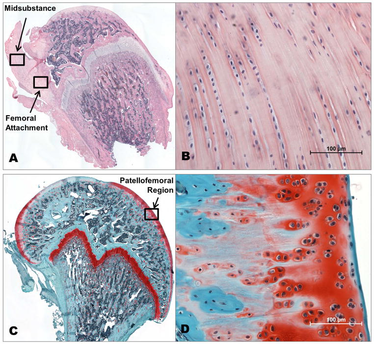
Administration of intra-articular medications, including corticosteroids and analgesics, is common clinical practice for knee pathology and dysfunction. Non-steroidal anti-inflammatory drugs (NSAIDs) are another category of medication commonly prescribed for their analgesic and anti-inflammatory properties. Recent studies demonstrated the efficacy of injectable NSAIDs in the treatment of intra-articular pathology and postoperative analgesia. However, little data exist regarding the safety of intra-articular injection, despite the increase in its application. Therefore, we investigated the effects of intra-articular NSAID injection on articular cartilage, the anterior cruciate ligament (ACL), and joint function in the rat. Sixty-four Sprague-Dawley rats were divided into either saline (SAL) or ketorolac (NSAID) tibiofemoral single injection treatment groups. Animals were euthanized at 2, 7, 28, and 84 days post-injection for histological and mechanical analyses. Additionally, a subset of animals underwent longitudinal ambulatory evaluation to determine joint functional properties. We hypothesized that intra-articular ketorolac injection would result in no detrimental mechanical, histological, or functional changes. No differences were reported between the NSAID and SAL groups in any of the parameters measured at any time point, demonstrating the potential safety of intra-articular NSAID administration. Therefore, NSAID injection could be further considered for clinical application in humans.
Keywords: NSAID; injection; intra-articular; knee; mechanics.
© 2014 Orthopaedic Research Society. Published by Wiley Periodicals, Inc.
Figures


Similar articles
-
Intra-articular injection of ketorolac in the rat knee joint: effect on articular cartilage and synovium.Br J Anaesth. 1998 Jun;80(6):837-9. doi: 10.1093/bja/80.6.837. Br J Anaesth. 1998. PMID: 9771318
-
Effect of Lubricin Mimetics on the Inhibition of Osteoarthritis in a Rat Anterior Cruciate Ligament Transection Model.Am J Sports Med. 2020 Mar;48(3):624-634. doi: 10.1177/0363546519898691. Epub 2020 Jan 31. Am J Sports Med. 2020. PMID: 32004084
-
Intra-Articular Injection of Cross-Linked Hyaluronic Acid-Dexamethasone Hydrogel Attenuates Osteoarthritis: An Experimental Study in a Rat Model of Osteoarthritis.Int J Mol Sci. 2016 Apr 15;17(4):411. doi: 10.3390/ijms17040411. Int J Mol Sci. 2016. PMID: 27092487 Free PMC article.
-
Knee kinematics, cartilage morphology, and osteoarthritis after ACL injury.Med Sci Sports Exerc. 2008 Feb;40(2):215-22. doi: 10.1249/mss.0b013e31815cbb0e. Med Sci Sports Exerc. 2008. PMID: 18202582 Review.
-
Reduction of systemic exposure and side effects by intra-articular injection of anti-inflammatory agents for osteoarthritis: what is the safer strategy?J Drug Target. 2023 Jul;31(6):596-611. doi: 10.1080/1061186X.2023.2220083. Epub 2023 Jun 9. J Drug Target. 2023. PMID: 37249274 Review.
Cited by
-
Effect of ketorolac in intra-articular injection analgesia for postoperative pain in patients undergoing shoulder arthroscopy: a pilot-controlled clinical study.J Pain Res. 2019 Jan 17;12:417-422. doi: 10.2147/JPR.S178413. eCollection 2019. J Pain Res. 2019. PMID: 30705607 Free PMC article.
-
Nonsteroidal Anti-Inflammatory Drug Injections versus Steroid Injections in the Management of Upper and Lower Extremity Orthopedic Conditions: A Systematic Review with Meta-Analysis.J Clin Med. 2024 Feb 17;13(4):1132. doi: 10.3390/jcm13041132. J Clin Med. 2024. PMID: 38398445 Free PMC article. Review.
-
Comparison of the effect of ketorolac versus triamcinolone acetonide injections for the treatment of de Quervain's tenosynovitis: a double-blind randomized controlled trial.BMC Musculoskelet Disord. 2022 Sep 1;23(1):831. doi: 10.1186/s12891-022-05784-x. BMC Musculoskelet Disord. 2022. PMID: 36050704 Free PMC article. Clinical Trial.
-
Pharmacokinetics, safety and efficacy of intra-articular non-steroidal anti-inflammatory drug injections for the treatment of osteoarthritis: A narrative review.J Clin Pharm Ther. 2022 Aug;47(8):1122-1133. doi: 10.1111/jcpt.13669. Epub 2022 May 3. J Clin Pharm Ther. 2022. PMID: 35505520 Free PMC article. Review.
-
The effects of intra-articular injection of ibuprofen on knee joint cartilage and synovium in rats.Acta Orthop Traumatol Turc. 2019 Jul;53(4):292-296. doi: 10.1016/j.aott.2019.03.013. Epub 2019 Apr 11. Acta Orthop Traumatol Turc. 2019. PMID: 30982756 Free PMC article.
References
-
- Convery PN, Milligan KR, Quinn P, et al. Low-dose intra-articular ketorolac for pain relief following arthroscopy of the knee joint. Anaesthesia. 1998;53:1125–1129. - PubMed
-
- Gupta A, Axelsson K, Allvin R, et al. Postoperative pain following knee arthroscopy: the effects of intra-articular ketorolac and/or morphine. Reg Anesth Pain Med. 1999;24:225–230. - PubMed
-
- Reuben SS, Connelly NR. Postoperative analgesia for outpatient arthroscopic knee sugery with intraarticular bupivacaine and ketorolac. Anesth Analg. 1995;80:1154–1157. - PubMed
-
- Praemer A, Furner S, Rice DP, et al. Musculoskeletal conditions in the United States. Park Ridge, Ill: American Academy of Orthopaedic Surgeons; 1992.
Publication types
MeSH terms
Substances
Grants and funding
LinkOut - more resources
Full Text Sources
Other Literature Sources
