Review
. 2014 Sep;10(9):504-16.
doi: 10.1038/nrneph.2014.112. Epub 2014 Jul 1.

Mechanisms of muscle wasting in chronic kidney disease

Affiliations
Review

Mechanisms of muscle wasting in chronic kidney disease

Xiaonan H Wang et al. Nat Rev Nephrol. 2014 Sep.

Abstract

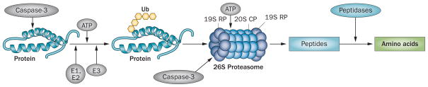
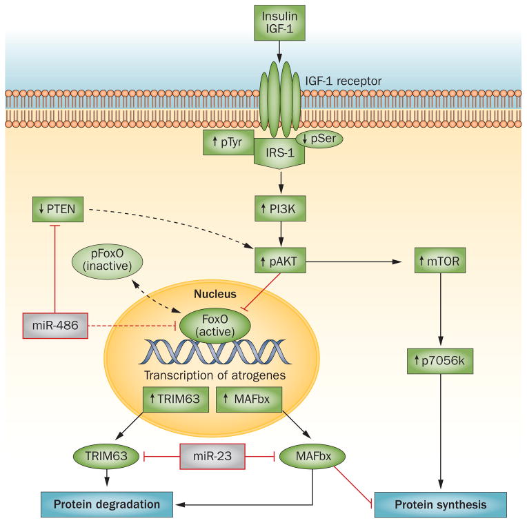
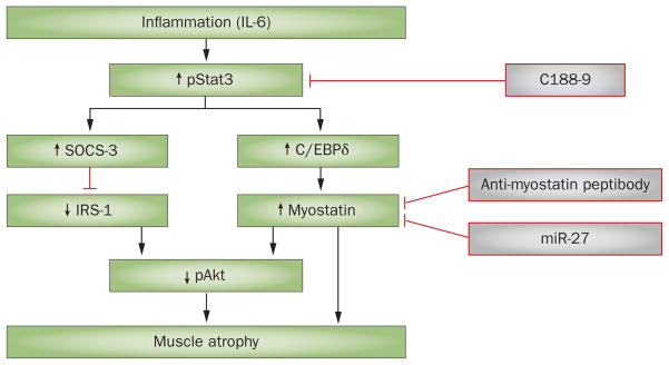
In patients with chronic kidney disease (CKD), loss of cellular proteins increases the risks of morbidity and mortality. Persistence of muscle protein catabolism in CKD results in striking losses of muscle proteins as whole-body protein turnover is great; even small but persistent imbalances between protein synthesis and degradation cause substantial protein loss. No reliable methods to prevent CKD-induced muscle wasting currently exist, but mechanisms that control cellular protein turnover have been identified, suggesting that therapeutic strategies will be developed to suppress or block protein loss. Catabolic pathways that cause protein wasting include activation of the ubiquitin-proteasome system (UPS), caspase-3, lysosomes and myostatin (a negative regulator of skeletal muscle growth). These pathways can be initiated by complications associated with CKD, such as metabolic acidosis, defective insulin signalling, inflammation, increased angiotensin II levels, abnormal appetite regulation and impaired microRNA responses. Inflammation stimulates cellular signalling pathways that activate myostatin, which accelerates UPS-mediated catabolism. Blocking this pathway can prevent loss of muscle proteins. Myostatin inhibition could yield new therapeutic directions for blocking muscle protein wasting in CKD or disorders associated with its complications.


Conflict of interest statement

Competing interests

The authors declare no competing interests.

Figures

Figure 1
Figure 1
Stages of skeletal muscle regeneration. Specific biomarkers can be detected at each stage of myogenesis. Satellite cells are normally quiescent, but when stimulated by factors such as insulin-like growth factor 1, they express myogenic regulatory factors, including myoblast determination protein 1. Expression of these factors leads to proliferation and differentiation of satellite cells into myoblasts and myocytes that can fuse to form myotubes or myofibrils. Abbreviations: Myf, myogenic factor; Pax, paired box protein.
Figure 2
Figure 2
Chronic kidney disease-induced protein degradation by the UPS. An initial step in the degradation cascade involves cleavage of the complex structure of muscle protein by caspase-3, which produces substrates for degradation. Protein substrates are conjugated to Ub by an ATP-dependent process involving the enzymes E1, E2 and E3. The selectivity of protein substrates principally depends on recognition of the protein to be degraded by specific E3 Ub-ligases (for example, TRIM63 for muscle proteins). After five Ub proteins are attached to the protein substrate, the complex can be recognized by the 26S proteasome, which releases Ubs, unfolds the protein substrate and ‘injects’ it into the 20S CP in which proteins are degraded to peptides. At this stage, caspase-3 also cleaves the 26S protease regulatory subunit 4 and 26S protease regulatory subunit 8, which are specific subunit proteins of the 19S proteasome RP. This reaction stimulates degradation of proteins in the 20S proteasome CP. Peptides released into the cytoplasm are degraded into amino acids. Abbreviations: CP, core particle; RP, regulatory particle; Ub, ubiquitin; UPS, ubiquitin–proteasome system.
Figure 3
Figure 3
Insulin/IGF-1 signalling stimulates protein synthesis and suppresses protein degradation. Binding of insulin or IGF-1 to the IGF-1 receptor stimulates tyrosine phosphorylation of IRS-1. Activation of IRS-1 stimulates the PI3K/Akt cascade, leading to phosphorylation of Akt, which activates mTOR (leading to protein synthesis) and phosphorylates (inactivates) Fox0. As pFoxO cannot enter the nucleus and, therefore, does not stimulate muscle protein losses, this process limits muscle protein wasting. In the absence of pAkt, active Fox0 increases transcription of the E3 ubiquitin ligases MAFbx and TRIM63, resulting in muscle wasting. MicroRNAs regulate IGF-1/PI3K/Akt signalling; miR-486 downregulates PTEN, which results in an increase in pAkt levels, whereas miR-23a suppresses translation of MAFbx and TRIM63 by interacting with their 3′-untranslated regions. The result is an inhibition of muscle atrophy. Abbreviations: FoxO, forkhead box protein O; IGF, insulin-like growth factor; IRS-1, insulin receptor substrate 1; MAFbx, F-box only protein 32; mTOR, mammalian target of rapamycin; p, phosphorylated; PI3K, phosphatidylinositol 3-kinase; PTEN, phosphatidylinositol 3,4,5-trisphosphate 3-phosphatase and dual-specificity protein phosphatase; TRIM63, E3 ubiquitin-protein ligase TRIM63.
Figure 4
Figure 4
A simplified mechanism of myostatin-induced muscle atrophy. Inflammation increases levels of circulating or local proinflammatory cytokines, such as IL-6. These cytokines stimulate phosphorylation of Stat3, which in turn stimulates SOCS-3 expression. The result is suppression of insulin/IGF-1 signalling, leading to muscle atrophy. IL-6-induced activation of pStat3 also stimulates C/EBPδ, which activates myostatin and, therefore, results in muscle loss. Activation of Stat3 can be blocked by the small-molecule inhibitor C188–9, which suppresses CKD-induced muscle wasting. Myostatin can also be blocked with either an anti-myostatin peptibody or miR-27 to prevent muscle wasting. Abbreviations: C/EBPδ, CCAAT/enhancer-binding protein δ; CKD, chronic kidney disease; IGF, insulin-like growth factor; IRS-1, insulin receptor substrate 1; SOCS-3, suppressor of cytokine signalling 3.

References

    1. Griffiths RD. Muscle mass, survival, and the elderly ICU patient. Nutrition. 1996;12:456–458. - PubMed
    1. Windsor JA, Hill GL. Risk factors for postoperative pneumonia. The importance of protein depletion. Ann Surg. 1988;208:209–214. - PMC - PubMed
    1. Gracia-Iguacel C, et al. Prevalence of protein-energy wasting syndrome and its association with mortality in haemodialysis patients in a centre in Spain. Nefrologia. 2013;33:495–505. - PubMed
    1. Go AS, Chertow GM, Fan D, McCulloch CE, Hsu CY. Chronic kidney disease and the risks of death, cardiovascular events, and hospitalization. N Engl J Med. 2004;351:1296–1305. - PubMed
    1. United States Renal Data System. USRDS 2009 Annual Data Report: Atlas of End-Stage Renal Disease in the United States. 2009 online http://www.usrds.org/atlas09.aspx.

Publication types

MeSH terms