. 2014 Jul 15;111(28):E2841-50.
doi: 10.1073/pnas.1401777111. Epub 2014 Jun 30.

Cep164 triggers ciliogenesis by recruiting Tau tubulin kinase 2 to the mother centriole

Affiliations

Cep164 triggers ciliogenesis by recruiting Tau tubulin kinase 2 to the mother centriole

Lukáš Čajánek et al. Proc Natl Acad Sci U S A. .

Abstract

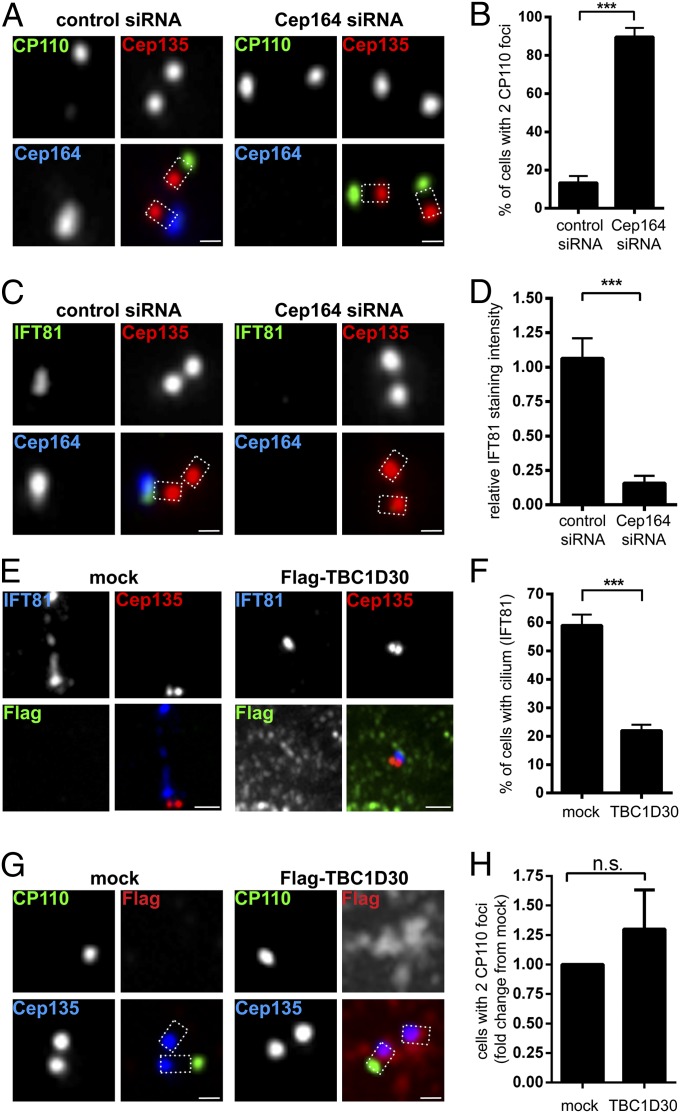
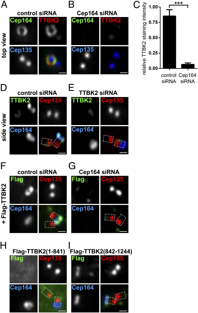
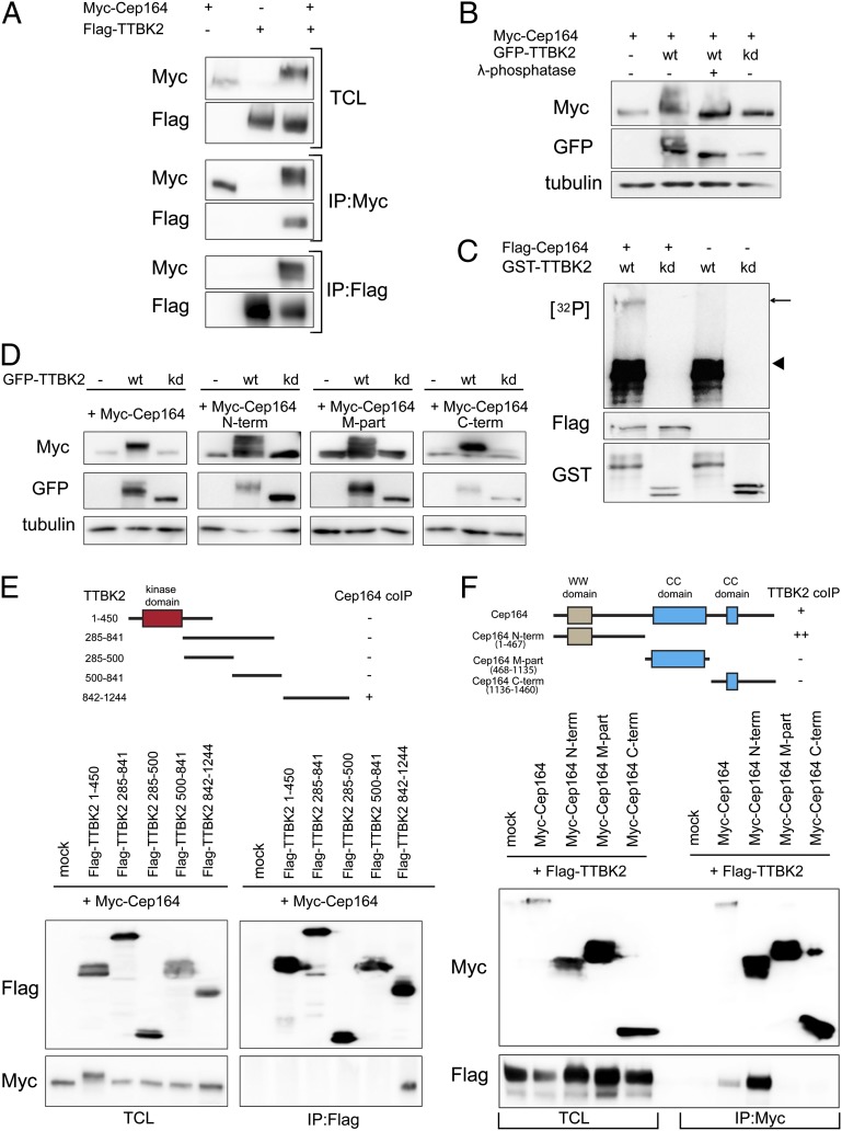
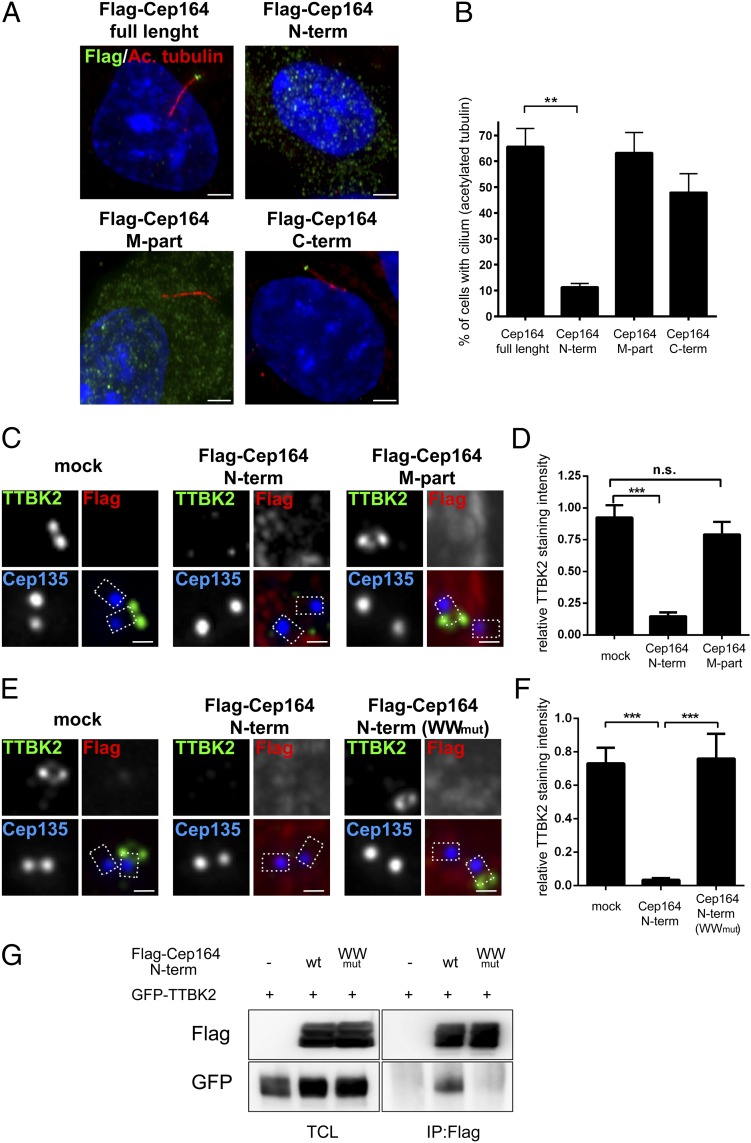
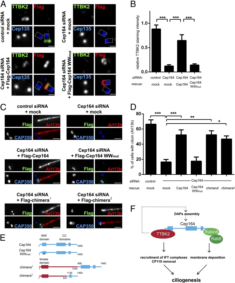
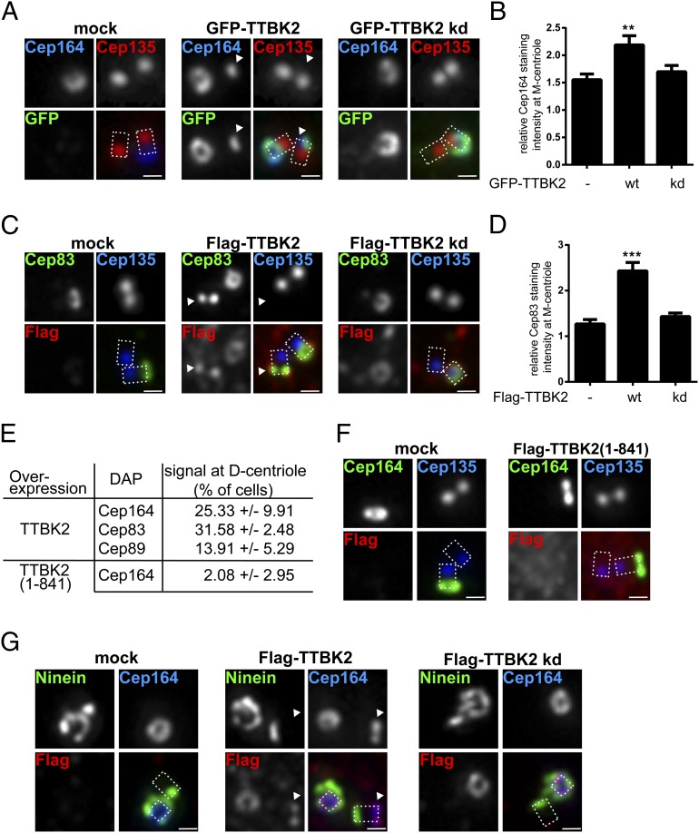
Primary cilia play critical roles in development and disease. Their assembly is triggered by mature centrioles (basal bodies) and requires centrosomal protein 164kDa (Cep164), a component of distal appendages. Here we show that loss of Cep164 leads to early defects in ciliogenesis, reminiscent of the phenotypic consequences of mutations in TTBK2 (Tau tubulin kinase 2). We identify Cep164 as a likely physiological substrate of TTBK2 and demonstrate that Cep164 and TTBK2 form a complex. We map the interaction domains and demonstrate that complex formation is crucial for the recruitment of TTBK2 to basal bodies. Remarkably, ciliogenesis can be restored in Cep164-depleted cells by expression of chimeric proteins in which TTBK2 is fused to the C-terminal centriole-targeting domain of Cep164. These findings indicate that one of the major functions of Cep164 in ciliogenesis is to recruit active TTBK2 to centrioles. Once positioned, TTBK2 then triggers key events required for ciliogenesis, including removal of CP110 and recruitment of intraflagellar transport proteins. In addition, our data suggest that TTBK2 also acts upstream of Cep164, contributing to the assembly of distal appendages.

Keywords: centrosome; primary cilium.


Conflict of interest statement

The authors declare no conflict of interest.

Figures

Fig. 1.
Fig. 1.
Depletion of Cep164 impairs early events in ciliogenesis independently of Rab8. RPE-1 cells were transfected with the indicated siRNA oligonucleotides (AD) or plasmids (EH) and serum starved. After fixation and permeabilization, cells were stained with the indicated antibodies and examined by indirect immunofluorescence microscopy. Cep135 staining was used to label proximal ends of centrioles. Dashed lines in merged images outline the centrioles. [Scale bars: 0.5 μm (A, C, and G) and 1 μm (E).] (A) Depletion of Cep164 from RPE-1 cells prevents removal of distal centriolar protein CP110 from the M centriole. (B) Quantification of the effects of Cep164 depletion on CP110 removal. Graph represents the summary of three experiments. (C) Loss of Cep164 impairs the recruitment of IFT81 to the M centriole. (D) Quantification of the effects of Cep164 depletion on IFT81 recruitment. Graph represents the summary of two experiments. (E) Transient expression of TBC1D30 (Rab8 GAP) impairs ciliogenesis, but not IFT81 localization to M centriole. (F) Quantification of the effects of TBC1D30 expression on ciliogenesis. Graph represents the summary of three experiments. (G) Transient expression of TBC1D30 does not prevent removal of CP110 from M centrioles. (H) Quantification of the effects of TBC1D30 expression on the CP110 removal. Graph represents the summary of four experiments. ***P < 0.001; n.s., not significant.
Fig. 2.
Fig. 2.
Cep164 is required for TTBK2 localization to the M centriole. Transfection and immunofluorescence experiments were carried out as described in the Fig. 1 legend. Cep135 staining was used to label proximal ends of centrioles. (Scale bars: 0.5 μm.) (A) Staining for endogenous Cep164 and TTBK2 reveals colocalization in a ring-like pattern at the M centriole, consistent with colocalization at distal appendages. (B) Depletion of Cep164 prevents recruitment of TTBK2 to the M centriole. (C) Quantification of the effects of Cep164 depletion on TTBK2 recruitment. Graph represents the summary of two experiments. ***P < 0.001. (D and E) Cep164 does not require TTBK2 for its localization to distal appendages of the M centriole. (F and G) Depletion of Cep164 prevents the recruitment of Flag-TTBK2 to the M centriole. (H) Flag–TTBK2 (1–841), lacking the Cep164 binding region (Fig. 3E), fails to localize to the M centriole. (I) Flag–TTBK2 (842–1244), containing the Cep164 binding region, localizes to the M centriole.
Fig. 3.
Fig. 3.
Cep164 is a binding partner and substrate of TTBK2. (A) HEK293T cells were transfected with the indicated constructs and subjected to immunoprecipitation (IP)–Western experiments. Upper shows total cell lysate (TCL). Western blots were performed by using the anti-tag antibodies listed to the left, and IPs were performed by using either anti-Myc (Myc–Cep164; Middle) or anti-Flag antibodies (Flag–TTBK2; Bottom). (B) Protein extracts, prepared from HEK293T cells transfected with Myc–Cep164 and GFP–TTBK [wild-type (WT) or kinase dead (kd) mutant], were treated with or without λ-phosphatase and analyzed by Western blotting. Note that Cep164 undergoes TTBK2-mediated phosphorylation, as indicated by a mobility shift that is sensitive to λ-phosphatase treatment. (C) In vitro-translated Flag–Cep164 was immunoprecipitated by using anti-Flag antibody and subjected to a kinase assay in the presence of [γ-32P]ATP and recombinant GST–TTBK2 (1-450) WT or kd protein. Arrow and arrowhead point to phosphorylated Flag–Cep164 and autophosphorylated GST–TTBK2 (1–450), respectively. (D) Protein extracts of transfected HEK293T cells were analyzed by Western blotting. Note that expression of GFP–TTBK2, but not kd mutant, led to prominently retarded electrophoretic mobility of Myc-tagged Cep164, Cep164 N term, Cep164 M part, and Cep164 C term. Also, note concomitant increases in levels of Cep164 and Cep164 C term. (E and F) Transfected HEK293T cells were subjected to IP–Western experiments. (Upper) Schematic representations of the constructs used are shown, and the symbols +, ++, or − summarize the efficacy of coimmunoprecipitation for each combination. (E) Domain mapping of TTBK2 identifies the C-terminal part (842–1244) as sufficient for Cep164 binding. (F) Domain mapping of Cep164 identifies the N-terminal part (1–467) as sufficient for interaction with TTBK2.
Fig. 4.
Fig. 4.
Excess of Cep164 N-terminal fragment impairs cilia formation by WW domain-mediated sequestration of TTBK2. Transfections and immunofluorescence experiments were carried out as outlined in the Fig. 1 legend. Cep135 staining was used to label proximal ends of centrioles. [Scale bars: 2.5 μm (A) and 0.5 μm (C and E).] (A) Localizations and impact on ciliogenesis of Cep164 full-length and deletion mutants (N term, 1–467; M part, 468–1135; C term, 1136–1460). PCs were visualized by staining for acetylated tubulin; note the dominant-negative effect of Cep164 N term. (B) Quantification of the data shown in A. Graph represents the summary of three experiments. (C) Expression of Cep164 N term, but not Cep164 M part, prevents TTBK2 localization to M centrioles. (D) Quantification of the data shown in C. Graph represents the summary of two experiments. (E) Expression of the WWmut mutant version of Cep164 N term (1–467) fails to impair M centriole localization of TTBK2. (F) Quantification of the data shown in E. Graph represents the summary of two experiments. (G) HEK293T cells were transfected with the indicated constructs and subjected to IP–Western experiments. Note that Cep164 N term (WWmut) fails to coimmunoprecipitate TTBK2. **P < 0.01; ***P < 0.001; n.s., not significant.
Fig. 5.
Fig. 5.
Tethering of TTBK2 to M centrioles rescues ciliogenesis in the absence of full-length Cep164. (AD) Transfections for siRNA-depletion/rescue experiments were carried out in RPE-1 cells, using the indicated siRNA oligonucleotides and rescue constructs; cells were then processed as outlined in the legend to Fig. 1. Cep135 staining visualizes proximal site of centrioles, and dashed lines in the merged images outline the centrioles. [Scale bars: 0.5 μm (A) and 1 μm (C).] (A) Expression of Cep164 full-length protein, but not the WWmut mutant, restores TTBK2 levels at M centrioles in Cep164-depleted cells. (B) Quantification of the rescue data shown in A. Graph represents the summary of three experiments. (C) Representative images illustrating the ability of the indicated constructs to rescue ciliogenesis. Staining for CAP350 and Arl13b was used to visualize centrioles and PC formation, respectively. (D) Quantification of the rescue of the rescue data shown in C. Graph represents the summary of at least four experiments. (E) Schematic representations of chimeric constructs used for siRNA-depletion/rescue experiments. Cep164 moieties are depicted in blue, and TTBK2 moieties are in red. (F) Model summarizing the crucial functions of TTBK2 and Cep164 domains in PC formation (see also Discussion). *P < 0.05; **P < 0.01; ***P < 0.001.
Fig. 6.
Fig. 6.
TTBK2 promotes assembly of DAPs at both M and D centrioles. Transfections of RPE-1 cells and immunofluorescence experiments were carried out as outlined in the Fig. 1 legend. Cep135 staining was used to label proximal ends of centrioles, and dashed lines in merged images outline the centrioles. (Scale bars: 0.5 μm.) Arrowheads point to distal parts of D centrioles. (A) Transient overexpression of GFP–TTBK2 promotes Cep164 localization to both M and D centrioles. (B) Quantification of the effects of TTBK2 overexpression on Cep164 levels at M centrioles. In cells where both centrioles showed Cep164 staining, the centriole showing more prominent Cep164 signal was considered as the M centriole. Graph represents the summary of two experiments. (C) Transient overexpression of Flag–TTBK2 promotes Cep83 localization to both M and D centrioles. (D) Quantification of the effects of TTBK2 overexpression on Cep83 levels at M centrioles. Graph represents the summary of two experiments. (E) Frequency of D centriole localizations of Cep164, Cep83, and Cep89 observed in transfected cells. Results were collected from at least two experiments. (F) Lack of influence of Flag–TTBK2 (1–841) overexpression on Cep164 localization. (G) Flag–TTBK2 promotes distal D centriole localization of Cep164 but not Ninein. Flag–TTBK2 kd is shown for control. **P < 0.01; ***P < 0.001.

References

    1. Singla V, Reiter JF. The primary cilium as the cell’s antenna: Signaling at a sensory organelle. Science. 2006;313(5787):629–633. - PubMed
    1. Goetz SC, Anderson KV. The primary cilium: A signalling centre during vertebrate development. Nat Rev Genet. 2010;11(5):331–344. - PMC - PubMed
    1. Gerdes JM, Davis EE, Katsanis N. The vertebrate primary cilium in development, homeostasis, and disease. Cell. 2009;137(1):32–45. - PMC - PubMed
    1. Satir P, Pedersen LB, Christensen ST. The primary cilium at a glance. J Cell Sci. 2010;123(Pt 4):499–503. - PMC - PubMed
    1. Ishikawa H, Marshall WF. Ciliogenesis: Building the cell’s antenna. Nat Rev Mol Cell Biol. 2011;12(4):222–234. - PubMed

Publication types

MeSH terms