. 2014 Jul 15;111(28):10335-40.
doi: 10.1073/pnas.1403286111. Epub 2014 Jun 30.

Primary cilia enhance kisspeptin receptor signaling on gonadotropin-releasing hormone neurons

Affiliations

Primary cilia enhance kisspeptin receptor signaling on gonadotropin-releasing hormone neurons

Andrew I Koemeter-Cox et al. Proc Natl Acad Sci U S A. .

Abstract

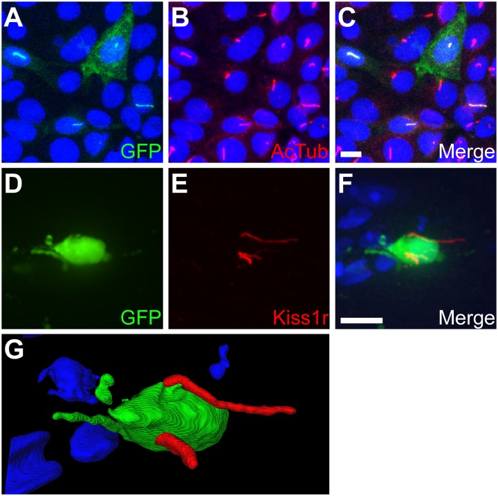
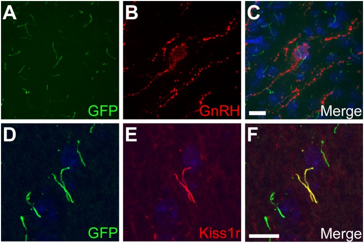
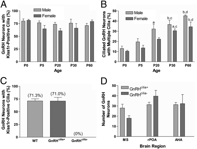
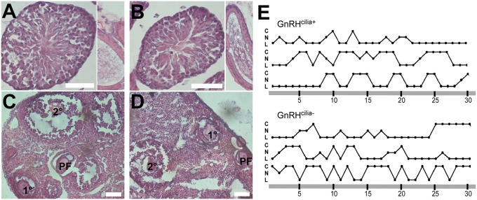
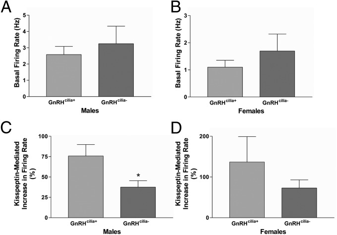
Most central neurons in the mammalian brain possess an appendage called a primary cilium that projects from the soma into the extracellular space. The importance of these organelles is highlighted by the fact that primary cilia dysfunction is associated with numerous neuropathologies, including hyperphagia-induced obesity, hypogonadism, and learning and memory deficits. Neuronal cilia are enriched for signaling molecules, including certain G protein-coupled receptors (GPCRs), suggesting that neuronal cilia sense and respond to neuromodulators in the extracellular space. However, the impact of cilia on signaling to central neurons has never been demonstrated. Here, we show that the kisspeptin receptor (Kiss1r), a GPCR that is activated by kisspeptin to regulate the onset of puberty and adult reproductive function, is enriched in cilia projecting from mouse gonadotropin-releasing hormone (GnRH) neurons. Interestingly, GnRH neurons in adult animals are multiciliated and the percentage of GnRH neurons possessing multiple Kiss1r-positive cilia increases during postnatal development in a progression that correlates with sexual maturation. Remarkably, disruption of cilia selectively on GnRH neurons leads to a significant reduction in kisspeptin-mediated GnRH neuronal activity. To our knowledge, this result is the first demonstration of cilia disruption affecting central neuronal activity and highlights the importance of cilia for proper GPCR signaling.

Keywords: GPR54; electrophysiology; neuronal primary cilia.


Conflict of interest statement

The authors declare no conflict of interest.

Figures

Fig. 1.
Fig. 1.
Kiss1r localizes to primary cilia in vitro and in vivo. (AC) Representative image of transiently transfected IMCD cells expressing Kiss1r fused at the C terminus to EGFP. (A) EGFP fluorescence (green) shows expression of Kiss1r. (B) Acetylated α-tubulin (AcTub; red) marks the cilia. (C) Merged image. (DF) Representative image of the medial hypothalamus in adult GnRH::GFP mice. (D) GFP fluorescence (green) indicates a GnRH neuron. (E) Labeling for Kiss1r (red) shows the presence of multiple Kiss1r-positive cilia. (F) Merged image. (G) 3D rendering of the same neuron confirms the cilia project from the same cell. Nuclei are stained with DRAQ5 (blue). (Scale bars: 10 µm.)
Fig. 2.
Fig. 2.
GnRH neurons possess cilia that are positive for Kiss1r. (AC) Representative image of the medial hypothalamus in adult CiliaGFP mice. (A) EGFP fluorescence (green) shows Sstr3 expression and ciliary localization. (B) Labeling for GnRH (red) indicates GnRH neurons. (C) Merged image confirms GnRH neurons are ciliated. (DF) Representative image of the medial hypothalamus in adult CiliaGFP mice. (D) EGFP fluorescence (green) shows Sstr3 expression and ciliary localization. (E) Labeling for Kiss1r (red) confirms Kiss1r ciliary localization. (F) Merged image. Nuclei are stained with DRAQ5 (blue). (Scale bars: 10 µm.)
Fig. 3.
Fig. 3.
Quantification of GnRH cilia. (A) Percentage of GnRH neurons with one or more Kiss1r-positive cilia in the medial hypothalamus of P0-P60 male and female GnRH::GFP mice (n = 4 animals of each sex at all ages, with the exception of P0, n = 3). Note the percentages do not vary significantly between ages or sexes. (B) Percentage of ciliated GnRH neurons with more than one Kiss1r-positive cilium in the medial hypothalamus of P0-P60 male and female GnRH::GFP mice (n = 4 animals of each sex at all ages, with the exception of P0, n = 3). Note the proportion of multiciliated GnRH neurons increases significantly between P0 and P60. aSignificantly different from P0 (P < 0.01). bSignificantly different from P0 (P < 0.001). cSignificantly different from P5 (P < 0.01). dSignificantly different from P5 (P < 0.001). (C) Percentage of GnRH neurons with one or more Kiss1r-positive cilia in the medial hypothalamus of P60 male GnRH::GFP, GnRHcilia+, and GnRHcilia- mice (n = 3–4 animals for each genotype). Note there is no difference in the percentage of Kiss1r-positive cilia between GnRH::GFP and GnRHcilia+ mice, but Kiss1r-positive cilia are completely lacking in GnRHcilia- mice. (D) Number of GnRH neurons throughout the medial septum (MS), rostral preoptic area (rPOA), and anterior hypothalamic area (AHA) of P60 male GnRHcilia+ and GnRHcilia- mice (n = 3 animals for each genotype). Note there is no significant difference in the number of GnRH neurons in any region between GnRHcilia+ and GnRHcilia- mice. Values are expressed as mean ± SEM.
Fig. 4.
Fig. 4.
GnRH cilia are dispensable for sexual maturation in male and female mice. (A and B) Representative testis (Left) and seminiferous tubule (Right) sections from P60 GnRHcilia+ (A) and GnRHcilia- (B) mice (n = 3 animals for each genotype) shows the presence of mature sperm in both genotypes. (C and D) Representative ovary section from P60 GnRHcilia+ (C) and GnRHcilia- (D) mice (n = 3 animals for each genotype) shows the presence of follicles at all stages of development. (Scale bars: 100 µm.) (E) Vaginal cytology of P60 GnRHcilia+ and GnRHcilia- mice show all stages of estrous cyclicity. C, cornified (estrous); L, leukocytic (metestrous and diestrous); N, nucleated (proestrous).
Fig. 5.
Fig. 5.
GnRH cilia are required for proper Kiss1r signaling. Basal firing rates of GnRH neurons from adult male (A) and ovariectomized female (B) GnRHcilia+ and GnRHcilia- mice. Note there is no significant difference in the basal firing rates between GnRHcilia+ and GnRHcilia- mice. Percentage increase in the firing rate of GnRH neurons after kisspeptin treatment in adult male (C) and ovariectomized female (D) GnRHcilia+ and GnRHcilia- mice. Note the increase in firing rate is significantly lower in male GnRHcilia- mice compared with GnRHcilia+ mice. Values are expressed as mean ± SEM. For males n = 10–13 neurons from 4 to 7 animals of each genotype. For females n = 9 neurons from 4 to 5 animals of each genotype. *Significantly different from GnRHcilia+ percentage (P = 0.02).

References

    1. Ishikawa H, Marshall WF. Ciliogenesis: Building the cell’s antenna. Nat Rev Mol Cell Biol. 2011;12(4):222–234. - PubMed
    1. Berbari NF, O’Connor AK, Haycraft CJ, Yoder BK. The primary cilium as a complex signaling center. Curr Biol. 2009;19(13):R526–R535. - PMC - PubMed
    1. Green JA, Mykytyn K. Neuronal ciliary signaling in homeostasis and disease. Cell Mol Life Sci. 2010;67(19):3287–3297. - PMC - PubMed
    1. Satir P, Pedersen LB, Christensen ST. The primary cilium at a glance. J Cell Sci. 2010;123(Pt 4):499–503. - PMC - PubMed
    1. Hildebrandt F, Benzing T, Katsanis N. Ciliopathies. N Engl J Med. 2011;364(16):1533–1543. - PMC - PubMed

Publication types

MeSH terms

LinkOut - more resources